Comparative Study
. 2011 Sep 6;50(35):7503-13.
doi: 10.1021/bi200826a. Epub 2011 Aug 16.

Schizosaccharomyces pombe protection of telomeres 1 utilizes alternate binding modes to accommodate different telomeric sequences

Affiliations
Comparative Study

Schizosaccharomyces pombe protection of telomeres 1 utilizes alternate binding modes to accommodate different telomeric sequences

Sarah E Altschuler et al. Biochemistry. .

Abstract

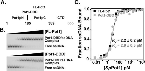
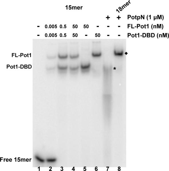
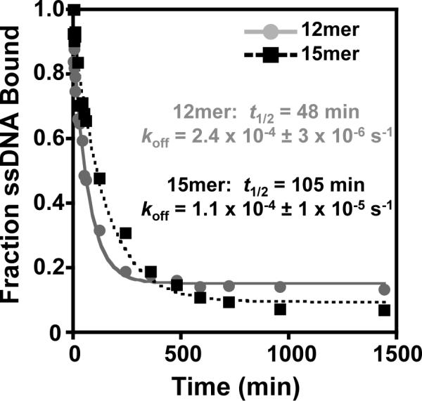
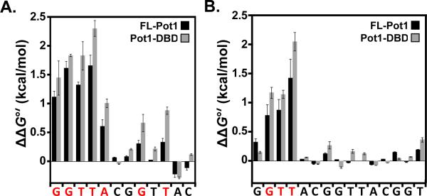
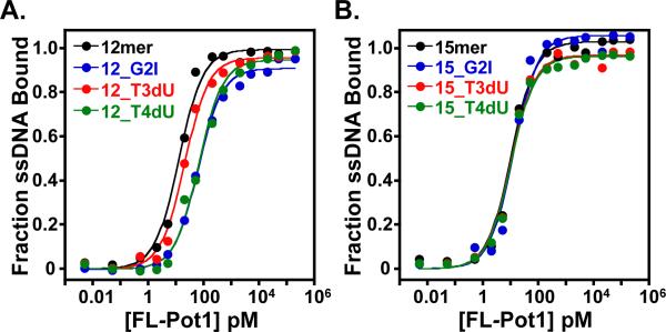
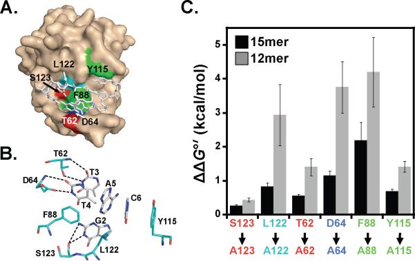
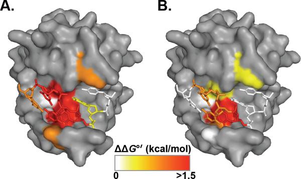
The ends of eukaryotic chromosomes consist of long tracts of repetitive GT-rich DNA with variable sequence homogeneity between and within organisms. Telomeres terminate in a conserved 3'-ssDNA overhang that, regardless of sequence variability, is specifically and tightly bound by proteins of the telomere-end protection family. The high affinity ssDNA-binding activity of S. pombe Pot1 protein (SpPot1) is conferred by a DNA-binding domain consisting of two subdomains, Pot1pN and Pot1pC. Previous work has shown that Pot1pN binds a single repeat of the core telomere sequence (GGTTAC) with exquisite specificity, while Pot1pC binds an extended sequence of nine nucleotides (GGTTACGGT) with modest specificity requirements. We find that full-length SpPot1 binds the composite 15mer, (GGTTAC)(2)GGT, and a shorter two-repeat 12mer, (GGTTAC)(2), with equally high affinity (<3 pM), but with substantially different kinetic and thermodynamic properties. The binding mode of the SpPot1/15mer complex is more stable than that of the 12mer complex, with a 2-fold longer half-life and increased tolerance to nucleotide and amino acid substitutions. Our data suggest that SpPot1 protection of heterogeneous telomeres is mediated through 5'-sequence recognition and the use of alternate binding modes to maintain high affinity interaction with the G-strand, while simultaneously discriminating against the complementary strand.


Figures

Figure 1
Figure 1
Domain organization and representative EMSA binding and analysis. (A) Domain boundaries are indicated for FL-Pot1, Pot1-DBD, Pot1pN, and Pot1pC. (B) Representative EMSA gels showing FL-Pot1 (top panel) and Pot1-DBD (lower panel) binding to 15mer ssDNA. (C) Normalized data were collected for triplicate EMSA experiments for FL-Pot1 (black squares) and Pot1-DBD (gray circles) binding to 15mer. Data are plotted as a function of fraction ssDNA bound versus Pot1 concentration (pM).
Figure 2
Figure 2
SpPot1 binds 15mer as a monomer. Increasing concentrations of a mixture of FL-Pot1 and Pot1-DBD (0, 0.005, 0.5, 50 nM; lanes 1–4) or 50 nM FL-Pot1 or Pot1-DBD alone (lanes 5–6) were added to 50 pM radiolabeled 15mer. For comparison, lane 7 shows 2× Pot1pN + 15mer (*) and lane 8 shows 3× Pot1pN + 18mer (◆). Total molecular weight of each complex: Pot1-DBD/15mer = 50 kDa; FL-Pot1/15mer = 69 kDa: Pot1pN/15mer = 50 kDa; Pot1pN/18mer = 73 kDa. The Pot1pN/15mer complex dissociates on the time-scale of the experiment.
Figure 3
Figure 3
The FL-Pot1/15mer complex displays a longer half-life than the FLPot1/12mer complex. FL-Potl/32 P-12mer complexes (grey circles) and FL-Pot1/32P-15mer complexes (black squares) were competed with a large molar excess of unlabeled 12mer or 15mer, respectively, (5 μM, 2500-fold excess over 32P-ssDNA) at various time points. Data are plotted as 32P-ssDNA bound versus time (min) and globally fit to a single-exponential decay model. Dissociation rate constant (koff) and half-life (t1/2) for each complex are indicated.
Figure 4
Figure 4
The specificity profiles determined by complementary base substitution in 12mer and 15mer on FL-Pot1 and Pot1-DBD reveal alternate modes of binding. (A) ΔΔG°' values for each 12mer complemer binding to FL-Pot1 (black bars) and Pot1-DBD (gray bars) were calculated as follows: ΔΔG°' = RT ln(KDcomplemer/KD12mer), where R = 1.9872 cal/(mol•K), T = 277.15 K, and KD12mer = 4.0 pM. (B) ΔΔG°' values for each 15mer complemer binding to FL-Pot1 (black bars) and Pot1-DBD (gray bars) were calculated as in (A): ΔΔG°' = RT ln(KDcomplemer/KD15mer), where R = 1.9872 cal/(mol•K), T = 277.15 K, and KD15mer = 2.2 pM.
Figure 5
Figure 5
Comparison of binding of non-canonical single nucleotide substituted ssDNA to FL-Pot1 reveals differential binding between 12mer and 15mer. (A) Binding curves for FL-Pot1 binding to 12mer (black) and the single non-canonical substitutions G2I (blue), T3dU (red) and T4dU (green) within 12mer. (B) Binding curves for FL-Pot1 binding to 15mer (black) and the single non-canonical substitutions G2I (blue), T3dU (red) and T4dU (green) within 15mer. Normalized data were collected for triplicate EMSA experiments and plotted as a function of fraction ssDNA bound versus FL-Pot1 concentration (pM).
Figure 6
Figure 6
Analysis of Pot1-DBD containing single alanine substitutions within ssDNA-binding interface of the N-terminal OB-fold reveals alternate binding modes for 12mer and 15mer. (A) Surface representation of Pot1pN/6mer crystal complex highlighting ssDNA-binding interface residues mutated to alanine. 6mer ssDNA ligand (sticks) is shown within the ssDNA-binding cleft for reference. (B) Hydrogen bonding and aromatic stacking interactions between protein residues (cyan) and nucleotides (white) within the ssDNA-binding interface. (C) ΔΔG°' values for 12mer (gray bars) and 15mer (black bars) binding to each Pot1-DBD alanine mutant were calculated as follows: ΔΔG°' = RT ln(KDala/KDwt), where R = 1.9872 cal/(mol•K), T = 277.15 K. Images in (A) and (B) were generated using PyMOL version 1.3 (53).
Figure 7
Figure 7
SpPot1 hot-spot of binding specificity. Representations of Pot1pN (surface) and bound 6mer (sticks) with the ssDNA-binding interface colored according to effects of substitution on 12mer (A) and 15mer (B) binding modes. ΔΔG°' < 0.6 kcal/mol (white), 0.6 – 1 kcal/mol (yellow), 1 – 1.5 kcal/mol (orange) > 1.5 kcal/mol (red). All images were generated using PyMOL version 1.3 (53).

References

    1. Croy J, Wuttke D. Themes in ssDNA recognition by telomereend protection proteins. Trends Biochem. Sci. 2006;31:516–525. - PubMed
    1. Bochkarev A, Bochkareva E. From RPA to BRCA2: lessons from single-stranded DNA binding by the OB-fold. Curr. Opin. Struct. Biol. 2004;14:36–42. - PubMed
    1. Ellenberger T, Tomkinson AE. Eukaryotic DNA ligases: structural and functional insights. Annu. Rev. Biochem. 2008;77:313–338. - PMC - PubMed
    1. Flynn RL, Zou L. Oligonucleotide/oligosaccharide-binding fold proteins: a growing family of genome guardians. Crit. Rev. Biochem. Mol. Biol. 2010;45:266–275. - PMC - PubMed
    1. Garvik B, Carson M, Hartwell L. Single-stranded DNA arising at telomeres in cdc13 mutants may constitute a specific signal for the RAD9 checkpoint. Mol. Cell. Biol. 1995;15:6128–6138. - PMC - PubMed

Publication types

MeSH terms

Substances