. 2011 Aug 4:5:351.
doi: 10.1186/1752-1947-5-351.

Large infrapatellar ganglionic cyst of the knee fat pad: a case report and review of the literature

Affiliations

Large infrapatellar ganglionic cyst of the knee fat pad: a case report and review of the literature

Ioannis Nikolopoulos et al. J Med Case Rep. .

Abstract

Introduction: Large ganglionic cystic formations arising from the infrapatellar fat pad are quite uncommon and only a few are mentioned in the literature. An open excision in these cases is mandatory.

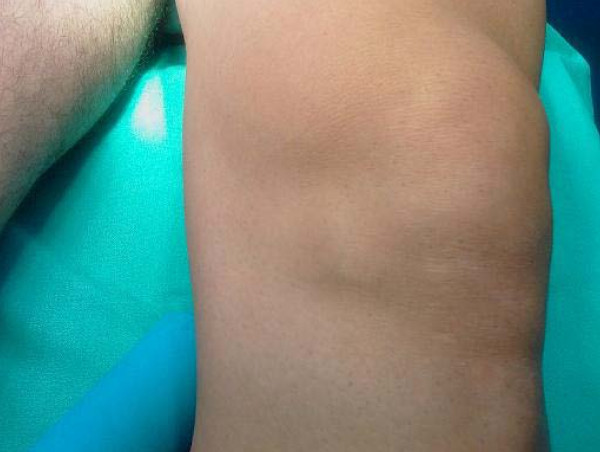
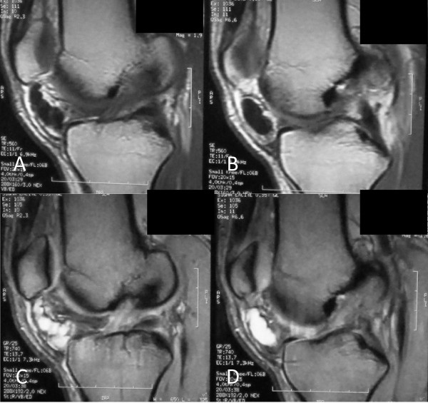
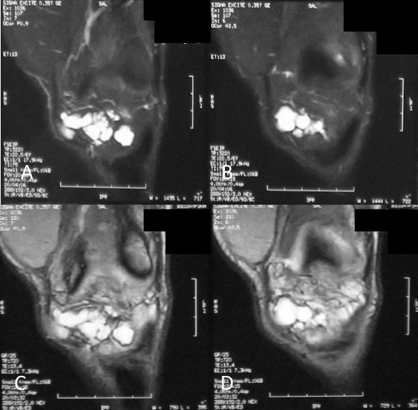
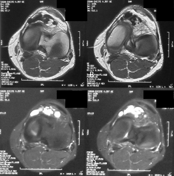
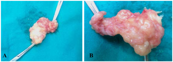
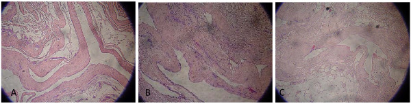
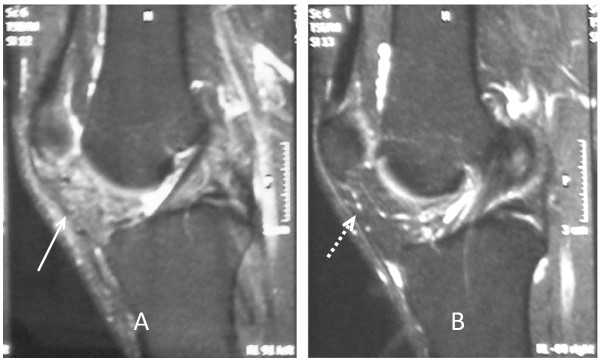
Case presentation: We report the case of a large infrapatellar fat pad ganglion in a 37-year-old Greek man with chronic knee discomfort. The ganglionic cyst originated from the infrapatellar fat pad and had no intrasynovial extension. The final diagnosis was determined with magnetic resonance imaging of the knee, and the lesion was treated with surgery.

Conclusions: These lesions are asymptomatic in most cases but often are misdiagnosed as meniscal or ligamentous lesions of the knee joint. Nowadays, the therapeutic trend for such lesions is arthroscopic excision, but when there is a large ganglion, as in this case report, the treatment should be an open and thorough resection. This report is intended mostly but not exclusively for clinical physicians and radiologists.


Figures

Figure 1
Figure 1
Swelling inferomedially to the patella of the left knee mimics a medial meniscal cyst.
Figure 2
Figure 2
Sagittal magnetic resonance images of the knee. (A, B) Sequential T1-weighted images of a cystic lesion with low signal intensity inferior to patella within the Hoffa's fat pad. (C, D) Sequential T2-weighted images, at the same level, of a well-demarcated, multilobular cystic mass with high signal intensity. Note the extrasynovial intra-articular location of the lesion.
Figure 3
Figure 3
Coronal magnetic resonance images of the anterior aspect of the knee. (A, B) Sequential T2-weighted images and (C, D) sequential proton density (PD) images of a multilobulated mass with high signal intensity.
Figure 4
Figure 4
Axial magnetic resonance images on the level of the inferior patellar pole. (A, B) Sequential T1-weighted images. (C, D) Sequential T2-weighted images.
Figure 5
Figure 5
(A) Cystic mass and incision site, (B) surgical approach and lesion exposure, and (C) synovium invasion and capsule defect after lesion excision.
Figure 6
Figure 6
(A) Intact total and (B) complete resected ganglionic cyst from Hoffa's fat pad with a portion of the inevitably invaded synovium.
Figure 7
Figure 7
Histologic sections of the specimen show the fibrous wall of the ganglion cyst with myxoid areas (A, B) and the presence of fatty cells adjacent to fibrous capsule (C) (hematoxylin and eosin [H&E], × 100).
Figure 8
Figure 8
Postoperative follow-up MRI of both knees. (A) Magnetic resonance imaging (MRI) of left knee six months after surgery shows the absence of cystic lesion with a mild increase of signal intensity of Hoffa's fat pad (arrow). (B) Right knee MRI shows a normal appearance and signal intensity of the infrapatellar fat pad (dashed arrow).

References

    1. Amin M, Torreggiani W, Sparks J. Infrapatellar ganglion that developed from infrapatellar fat and had minimal intraarticular extension. Knee Surg Sports Traumatol Arthrosc. 2008;16:179–181. doi: 10.1007/s00167-007-0406-9. - DOI - PubMed
    1. Antonacci V, Foster T, Fenlon H, Harper K, Eustace S. Technical report: CT-guided aspiration of anterior cruciate ligament ganglion cysts. Clin Radiol. 1998;53:771–773. doi: 10.1016/S0009-9260(98)80323-1. - DOI - PubMed
    1. Bui-Mansfield LT, Youngberg RA. Intraarticular ganglia of the knee: prevelance, presentation, etiology and management. Am J Roentgenol. 1997;168:123–127. - PubMed
    1. Kaempffe F, D'Amato C. An unusual intra-articular ganglion of the knee with interosseous extension; a case report. J Bone Joint Surg Am. 1989;71:773–775. - PubMed
    1. Feldman F, Singson DR, Staron BR. Magnetic resonance imaging of paraarticular and ectopic ganglia. Skeletal Radiol. 1989;18:353–358. doi: 10.1007/BF00361424. - DOI - PubMed

LinkOut - more resources