Review
. 2011 Aug 4:10:63.
doi: 10.1186/1475-2859-10-63.

Polyphosphate--an ancient energy source and active metabolic regulator

Affiliations
Review

Polyphosphate--an ancient energy source and active metabolic regulator

Lucia Achbergerová et al. Microb Cell Fact. .

Abstract

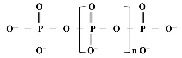
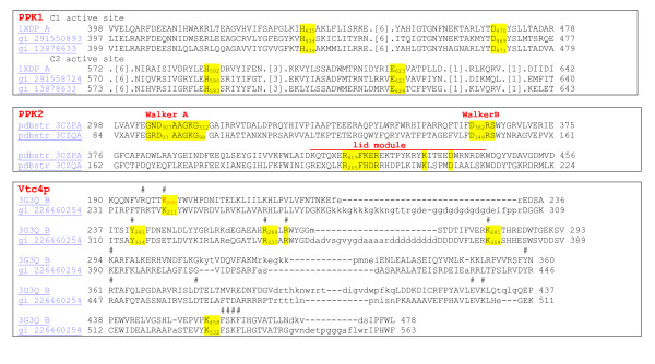
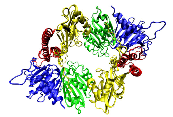
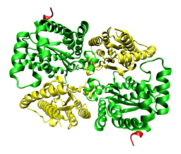
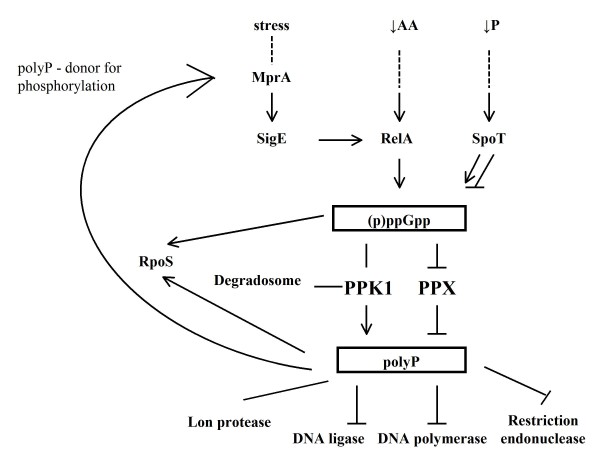
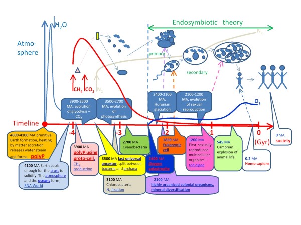
There are a several molecules on Earth that effectively store energy within their covalent bonds, and one of these energy-rich molecules is polyphosphate. In microbial cells, polyphosphate granules are synthesised for both energy and phosphate storage and are degraded to produce nucleotide triphosphate or phosphate. Energy released from these energetic carriers is used by the cell for production of all vital molecules such as amino acids, nucleobases, sugars and lipids. Polyphosphate chains directly regulate some processes in the cell and are used as phosphate donors in gene regulation. These two processes, energetic metabolism and regulation, are orchestrated by polyphosphate kinases. Polyphosphate kinases (PPKs) can currently be categorized into three groups (PPK1, PPK2 and PPK3) according their functionality; they can also be divided into three groups according their homology (EcPPK1, PaPPK2 and ScVTC). This review discusses historical information, similarities and differences, biochemical characteristics, roles in stress response regulation and possible applications in the biotechnology industry of these enzymes. At the end of the review, a hypothesis is discussed in view of synthetic biology applications that states polyphosphate and calcium-rich organelles have endosymbiotic origins from ancient protocells that metabolized polyphosphate.


Figures

Figure 1
Figure 1
The polyphosphate molecule.
Figure 2
Figure 2
Schematic diagram showing the key catalytic residues of PPK1, PPK2, and VTC4.
Figure 3
Figure 3
Crystal structure of E. coli asymmetric unit polyphosphate kinase 1 [62]and its implication for polyphosphate synthesis. PPK1 contains two asymmetric units. On the picture, there is an asymmetric unit of PPK1, which contains two monomers. Each monomer of PPK1 contains amino-terminal domain (N domain) coloured in red, the "head" domain (H domain) in yellow and two carboxyterminal domains (C1 and C2 domains) in green and blue. The coordinates were downloaded from Protein Data Bank (the corresponding PDB code- 1XDO) [63] and visualized by Visual Molecular Dynamics 1.9 [64] and POV-Ray [65].
Figure 4
Figure 4
Crystal structures of P. aeruginosa polyphosphate kinase 2 [60]and their implications for polyphosphate synthesis. P. aeruginosa PPK2 contains two monomers. Each monomer contains two domains coloured in yellow and green connected by a flexible linker coloured in red. The coordinates were downloaded from Protein Data Bank (the corresponding PDB codes- 3CZP) [63] and visualized by Visual Molecular Dynamics 1.9 [64] and POV-Ray [65].
Figure 5
Figure 5
Crystal structures of Sinorhizobium meliloti polyphosphate kinase 2 [60]and their implications for polyphosphate synthesis. S. meliloti PPK2 contains four monomers. Each monomer contains two domains coloured in yellow and green connected by a flexible linker coloured in red. The coordinates were downloaded from Protein Data Bank (the corresponding PDB codes- 3CZQ) [63] and visualized by Visual Molecular Dynamics 1.9 [64] and POV-Ray [65].
Figure 6
Figure 6
Crystal structure of Saccharomyces cerevisiae VTC4 [46]and its implication for polyphosphate synthesis. VTC4 contains two monomers coloured in red and blue. The coordinates were downloaded from Protein Data Bank (the corresponding PDB code- 3G3Q) [63] and visualized by Visual Molecular Dynamics 1.9 [64] and POV-Ray [65].
Figure 7
Figure 7
The cenral role of PPK1 in metabolism involved in gene and protein regulation. ↓AA - amino acid starvation; ↓P - phosphate starvation; (p)ppGpp -guanosine (penta)tetraphosphate; → activation; ⊥ inhibition.
Figure 8
Figure 8
Timeline of key events in the global history of life evolution, integrating evidence for the endosymbiotic theory and insertion of the hypothesis of polyP-protocells. This scheme was designed to depict synthetic biology section. MA - million years ago.

References

    1. Lodish H. Molecular cell biology. 6. New York: W.H. Freeman; 2007.
    1. Kulaev IS. Biochemistry of inorganic polyphosphates. Rev Physiol Biochem Pharmacol. 1975;73:131–158. doi: 10.1007/BFb0034661. - DOI - PubMed
    1. Yamagata Y, Watanabe H, Saitoh M, Namba T. Volcanic production of polyphosphates and its relevance to prebiotic evolution. Nature. 1991;352:516–519. doi: 10.1038/352516a0. - DOI - PubMed
    1. Gower LB. Biomimetic model systems for investigating the amorphous precursor pathway and its role in biomineralization. Chem Rev. 2008;108:4551–4627. doi: 10.1021/cr800443h. - DOI - PMC - PubMed
    1. Diaz J, Ingall E, Benitez-Nelson C, Paterson D, de Jonge MD, McNulty I, Brandes JA. Marine polyphosphate: a key player in geologic phosphorus sequestration. Science. 2008;320:652–655. doi: 10.1126/science.1151751. - DOI - PubMed

Publication types

MeSH terms

Substances

LinkOut - more resources