Review
. 2011 Aug 5:12:33.
doi: 10.1186/1471-2199-12-33.

Cruciform structures are a common DNA feature important for regulating biological processes

Affiliations
Review

Cruciform structures are a common DNA feature important for regulating biological processes

Václav Brázda et al. BMC Mol Biol. .

Abstract

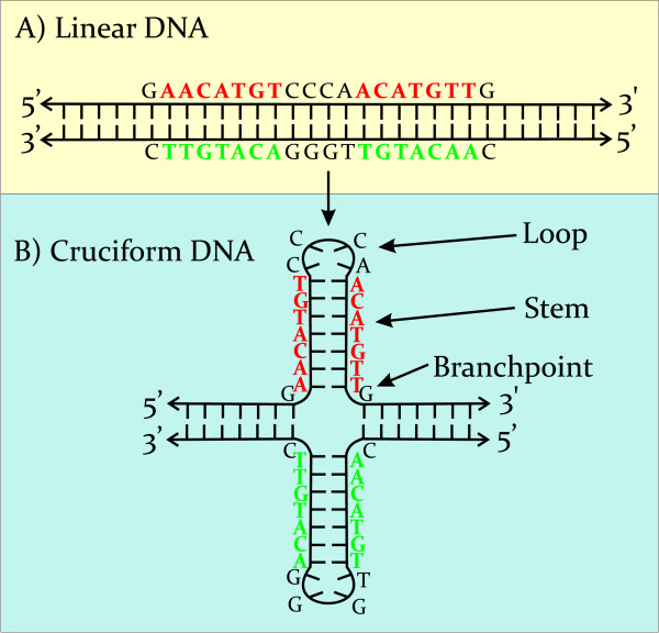
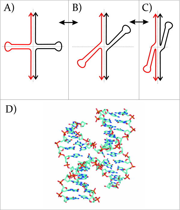
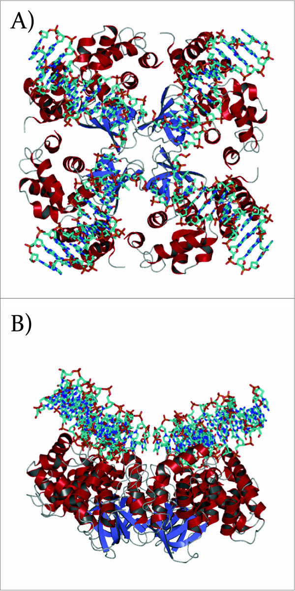
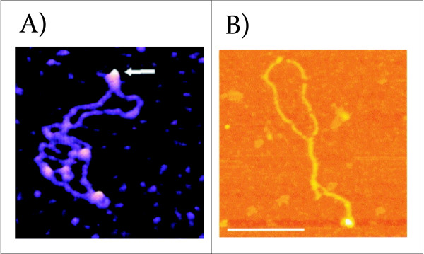
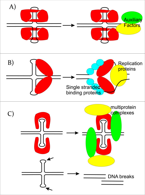
DNA cruciforms play an important role in the regulation of natural processes involving DNA. These structures are formed by inverted repeats, and their stability is enhanced by DNA supercoiling. Cruciform structures are fundamentally important for a wide range of biological processes, including replication, regulation of gene expression, nucleosome structure and recombination. They also have been implicated in the evolution and development of diseases including cancer, Werner's syndrome and others.Cruciform structures are targets for many architectural and regulatory proteins, such as histones H1 and H5, topoisomerase IIβ, HMG proteins, HU, p53, the proto-oncogene protein DEK and others. A number of DNA-binding proteins, such as the HMGB-box family members, Rad54, BRCA1 protein, as well as PARP-1 polymerase, possess weak sequence specific DNA binding yet bind preferentially to cruciform structures. Some of these proteins are, in fact, capable of inducing the formation of cruciform structures upon DNA binding. In this article, we review the protein families that are involved in interacting with and regulating cruciform structures, including (a) the junction-resolving enzymes, (b) DNA repair proteins and transcription factors, (c) proteins involved in replication and (d) chromatin-associated proteins. The prevalence of cruciform structures and their roles in protein interactions, epigenetic regulation and the maintenance of cell homeostasis are also discussed.


Figures

Figure 1
Figure 1
Changes associated with transition from the linear to cruciform state in the p53 target sequence from the p21 promoter. The promoter sequence contains a 20 bp p53 target sequence with 7 bp long inverted repeat (red), (A) as linear DNA and (B) as an inverted repeat as a cruciform structure. In the cruciform structure, the p53 target sequence is presented as stems and loops.
Figure 2
Figure 2
Conformations of a cruciform structure. Conformations of a cruciform can vary from (A) "unfolded" with 4-fold symmetry to (B) bent, and to (C) "stacked" with 4 chains of DNA in close vicinity. D) Topology of a Holliday junction stabilized by a psoralen cross-linking agent (PDBID 467D). Here, the junction takes the form of an anti-parallel stacked x-structure.
Figure 3
Figure 3
Crystal structure of the E. coli RuvA tetramer in complex with a Holliday junction (PDBID 1C7Y). A) The Holliday junction is depressed at the center where it makes close contacts with RuvA. Each of the arms outside of the junction center takes on a standard beta-DNA conformation B) Rotation of A) by 90°.
Figure 4
Figure 4
AFM and SFM images of proteins binding to a cruciform structure. A) AFM images of PARP-1 binding to supercoiled pUC8F14 plasmid DNA containing a 106 bp inverted repeat. PARP-1 binds to the end of the hairpin arm (white arrow). Images show 300 × 300 nm2 surface areas (reprinted with permission from [51]. B) The interaction between p53CD and supercoiled DNA gives rise to cruciform structures. Shown is an SFM image of complex formed between p53CD and sc pXG(AT)34 plasmid DNA at a molar ratio of 2.5; the complexes were mounted in the presence of 10 mM MgAc2. The scale bars represent 200 nm (reprinted with permission from [132].
Figure 5
Figure 5
Scheme of the putative mechanistic roles of cruciform binding proteins in transcription, DNA replication, and DNA repair. A) A model for the structure-specific binding of transcription factors to a cognate palindrome-type cruciform implicated in transcription. The equilibrium between classic B-DNA and the higher order cruciform favors duplex DNA, but, when cruciform binding proteins are present, they either preferentially bind to and stabilize the cruciform or bind to the classic form and convert it to the cruciform. This interaction results in both an initial melting of the DNA region covered by transcription factor and an extension of the melt region in both directions. The melting region continues to extend in response to the needs of the active transcription machinery. B) A model for the initiation of replication enhanced by extrusion to a cruciform structure. Dimeric cruciform binding proteins interact with and stabilize the cruciform structure. The replisome is assembled concomitantly and is assumed to include polymerases, single-strand binding proteins and helicases. C) Model for the influence of cruciform binding proteins on DNA structure in DNA damage regulation. Naked cruciforms are sensitive to DNA damage and are covered by proteins in order to protect these sequences from being cleaved. In these cases, a deficiency in cruciform binding proteins can lead to DNA breaks. Here, cruciform-DNA complexes can also serve as scaffolds to recruit the DNA damage machinery.

References

    1. Smith GR. Meeting DNA palindromes head-to-head. Genes Dev. 2008;22(19):2612–2620. doi: 10.1101/gad.1724708. - DOI - PMC - PubMed
    1. Palecek E. Local supercoil-stabilized DNA structures. Crit Rev Biochem Mol Biol. 1991;26:151–226. doi: 10.3109/10409239109081126. - DOI - PubMed
    1. van Holde K, Zlatanova J. Unusual DNA structures, chromatin and transcription. Bioessays. 1994;16(1):59–68. doi: 10.1002/bies.950160110. - DOI - PubMed
    1. Krasilnikov AS, Podtelezhnikov A, Vologodskii A, Mirkin SM. Large-scale effects of transcriptional DNA supercoiling in vivo. J Mol Biol. 1999;292(5):1149–1160. doi: 10.1006/jmbi.1999.3117. - DOI - PubMed
    1. Mikheikin AL, Lushnikov AY, Lyubchenko YL. Effect of DNA supercoiling on the geometry of holliday junctions. Biochemistry. 2006;45(43):12998–13006. doi: 10.1021/bi061002k. - DOI - PMC - PubMed

Publication types