. 2011 Aug 5;146(3):396-407.
doi: 10.1016/j.cell.2011.06.042.

The Min oscillator uses MinD-dependent conformational changes in MinE to spatially regulate cytokinesis

Affiliations

The Min oscillator uses MinD-dependent conformational changes in MinE to spatially regulate cytokinesis

Kyung-Tae Park et al. Cell. .

Abstract

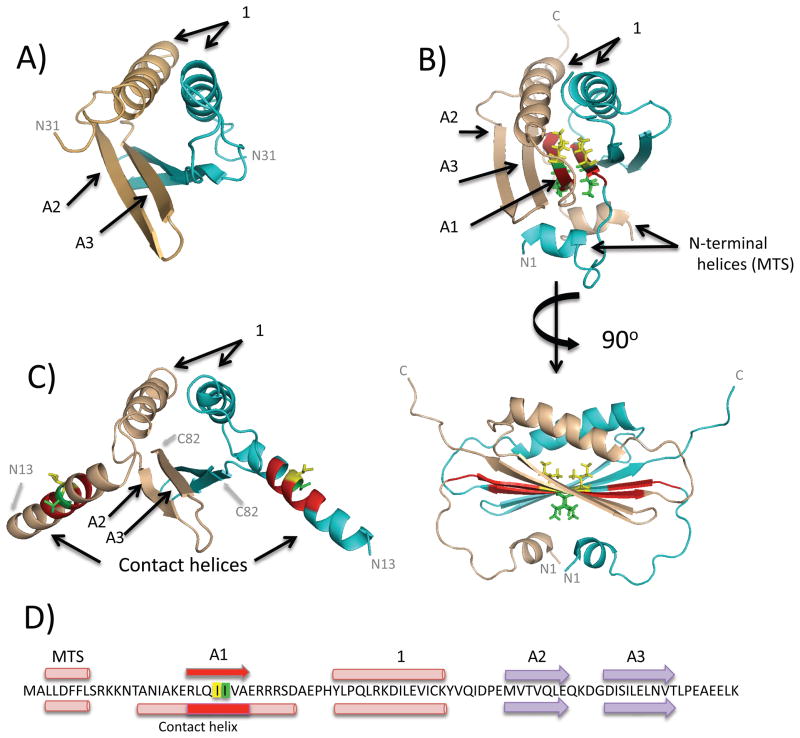
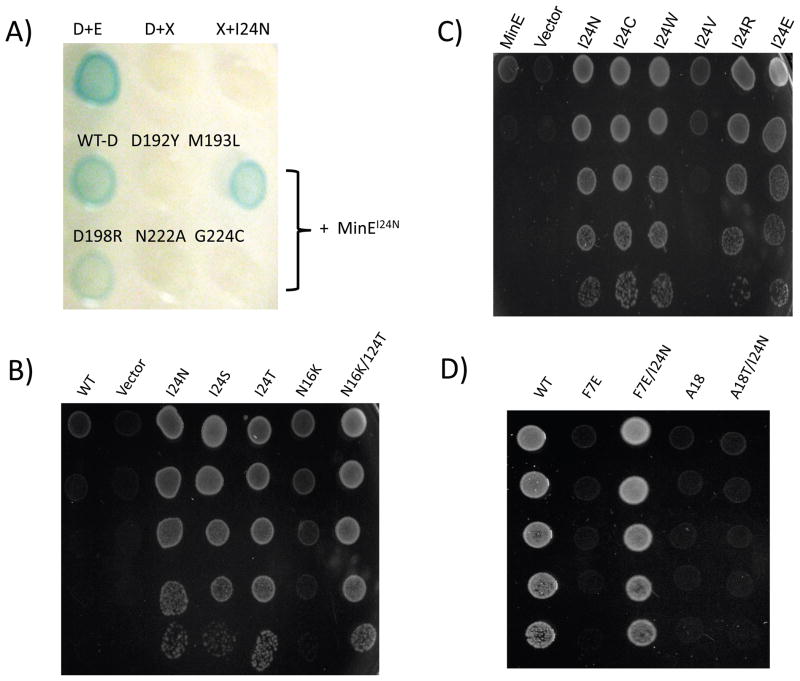
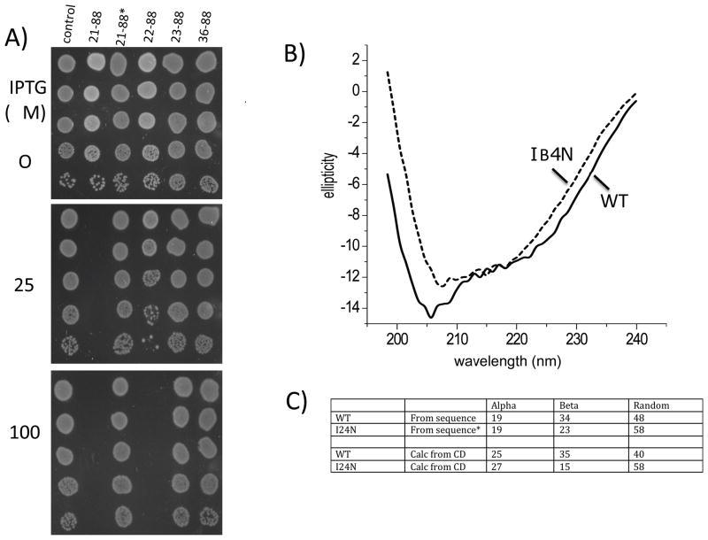
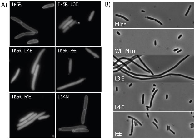
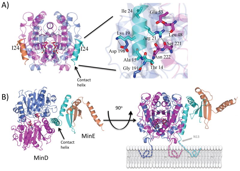
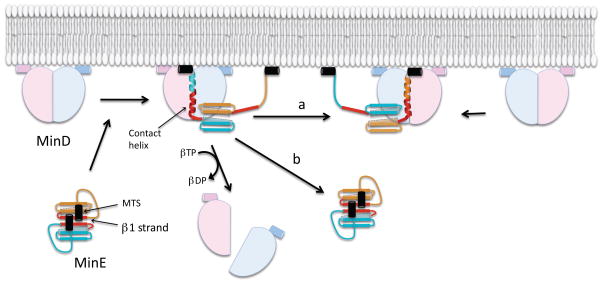
In E. coli, MinD recruits MinE to the membrane, leading to a coupled oscillation required for spatial regulation of the cytokinetic Z ring. How these proteins interact, however, is not clear because the MinD-binding regions of MinE are sequestered within a six-stranded β sheet and masked by N-terminal helices. minE mutations that restore interaction between some MinD and MinE mutants were isolated. These mutations alter the MinE structure leading to release of the MinD-binding regions and the N-terminal helices that bind the membrane. Crystallization of MinD-MinE complexes revealed a four-stranded β sheet MinE dimer with the released β strands (MinD-binding regions) converted to α helices bound to MinD dimers. These results identify the MinD-dependent conformational changes in MinE that convert it from a latent to an active form and lead to a model of how MinE persists at the MinD-membrane surface.


Figures

Fig. 1
Fig. 1
Structures of MinE and location of critical residues. The structure of the trypsin-treated MinE (residues 31–88) from E. coli (A) and two views of the the MinE (residues 1–89) from N. gonnorrhoeae (B) are shown (PDBs 1EVO and 2KXO, respectively). These structures contain 4-stranded and 6-stranded β-sheets, respectively. The labeling of secondary structural elements follows the labeling of the N. gonnorrhoeae structure. The N-terminal helices are shown in this study to function as a membrane targeting sequence (MTS). The residue corresponding to I24 of the E. coli MinE is colored yellow in this structure and the residue corresponding to I25 is colored green. (C) The structure of MinE12–88(I24N) from the MinD-MinE complex reported in this work (residues 13–83 of MinE are visible). Note that it is a 4-stranded β-sheet and the region corresponding to β1 in panel B (red) is part of an α helix (the contact helix). The N- and C-termini are indicated. (D) The sequence of MinE from E. coli with the secondary structural elements present in free MinE displayed above the sequence and those present in MinE in the complex with MinD displayed below the sequence.
Fig. 2
Fig. 2
Analysis of the ability of MinE mutants to bind to MinD mutants and suppress MinC/MinD inhibitory activity. A) Bacterial two-hybrid analysis of the interaction between MinEI24N and several MinD mutants. First row (controls): MinD + MinE, MinD + X and X + MinEI24N (X=empty vector); the second and third row contain MinEI24N in combination with the indicated MinD mutant. B) The ability of various minE alleles to suppress killing by MinC/MinDM193L. JS964 (Δmin)/pSEB104CD-193 (Para::minC minDM193L) with pJB216 (Plac::minE) derivatives containing the indicated minE allele were serially diluted 10 fold and spotted on plates containing 0.1% arabinose and 100 μM IPTG. C) As in panel B. D) The minEI24N mutation suppresses some, but not all, minE mutations. pJB216 (Plac::minE) derivatives carrying the minE alleles indicated were tested for their ability to protect JS964 (Δmin) from the induction of MinC/MinD from pSEB104CD (Para::minC minD).
Fig. 3
Fig. 3
Inhibitory activity and secondary structure of N-terminal truncated MinEs. A) The sensitivity of JS219 (min+) to N-terminally truncated MinEs was determined by spotting serial (10-fold) dilutions of cultures of JS219 containing plasmids expressing various N-terminal truncated MinE derivatives on plates containing IPTG as indicated. The control is the parent vector without an insert. The presence of the I24N substitution is indicated by the asterisk. B) Circular dichroism spectra of MinE21–88 and MinE21–88(I24N). C) The % of secondary structure content was estimated from the CD spectra using the K2d prediction program (Andrade et al., 1993) and is compared to the % of secondary structure content present in the crystal structure of MinE (corresponding to residues 21–88) from H. pylori. The asterisk indicates that the value for β content of MinE21–88(I24N) was calculated assuming the β1 strand is a random coil.
Fig. 4
Fig. 4
Effect of minE mutations on membrane localization of MinE and its ability to counter MinC/MinD. A) JS964 (Δmin) containing pJK100 (Ptrc::minE-gfp) derivatives expressing minE-GFP fusions with the indicated minE mutations were analyzed by fluorescence microscopy. The strains were grown in the presence of 20 μM IPTG. B) The effect of minE mutations on spatial regulation of cell division. JS964 (Δmin) containing pSEB104CDE (Para::minC minD minE) derivatives containing various minE mutations (as indicated in the panels) was grown to exponential phase with 0.1% arabinose for 24 hours to induce the min operon. The first panel contained the minCR133A mutation, which prevents interaction with MinD and inactivates Min function (Zhou and Lutkenhaus, 2005).
Fig. 5
Fig. 5
Structure of the MinE-MinD complex. Panel (A) contains the complex between MinDΔ10D40A and MinE12–31 (only residues 13–26 are visible). The structure shows a MinE peptide (contact helix; colored cyan and organe) bound to each side of a MinD dimer (magenta and blue: ADP in red). On the right is a blowup of the MinE contact helix bound to MinD. It is rotated 90°. Hydrogen bonds are indicated by dashed lines. The I24 residue is on the side of the helix away from MinD. Panel (B) shows the structure of the complex between MinDΔ10D40A and MinEI24N*-h (both dimers). In the crystal the dimers alternate to make a continuous helix (Fig. S5C). In the orientation on the left the membrane binding surface of MinD is beneath MinD so that the N-terminus of the contact helix (residue 13) is directed into the plane of the figure. In the structure on the right the MinD-MinE complex is rotated 90° so the orientation with respect to the membrane can be observed. The MTSs of MinD and MinE are depicted with dotted lines.
Fig. 6
Fig. 6
Tarzan of the Jungle model for the interaction between MinD and MinE. In this model MinE encounters MinD bound to the membrane and the MTSs (black segments) and the β1 strands (red) of MinE are released from the 6 stranded β-sheet structure resulting in formation of a 4-stranded β-sheet structure. One of the released β1 strands along with N-terminal flanking residues form an α-helix that is stabilized by binding to MinD while the other is tethered to the membrane through its linked MTS. The fate of MinE depends on two competing reactions (indicated by ‘a’ and ‘b’ ) following the dissociation of MinD due to ATPase stimulation. Either it is handed off to another MinD (a) or dissociates from the membrane as it snaps back to the 6 β-stranded structure (b). A higher density of MinD on the membrane favors the former.

References

    1. Andrade MA, Chacon P, Merelo JJ, Moran F. Evaluation of secondary structure of proteins from UV circular dichroism spectra using an unsupervised learning neural network. Protein Eng. 1993;6:383–390. - PubMed
    1. Arjunan SN, Tomita S. A new multicompartmental reaction-diffusion modeling method links transient membrane attachment of E. coli MinE to E-ring formation. Syst Synth Biol. 2010;4:35–53. - PMC - PubMed
    1. Barilla D, Carmelo E, Hayes F. The tail of the ParG DNA segregation protein remodels ParF polymers and enhances ATP hydrolysis via an arginine finger-like motif. Proc Natl Acad Sci U S A. 2007;104:1811–1816. - PMC - PubMed
    1. de Boer PA, Crossley RE, Rothfield LI. A division inhibitor and a topological specificity factor coded for by the minicell locus determine proper placement of the division septum in E. coli. Cell. 1989;56:641–649. - PubMed
    1. Evans P. Scaling and assessment of data quality. Acta Crystallogr D Biol Crystallogr. 2006;62:72–82. - PubMed

Publication types

MeSH terms