. 2011 Aug 5;9(2):144-55.
doi: 10.1016/j.stem.2011.06.015.

BRACHYURY and CDX2 mediate BMP-induced differentiation of human and mouse pluripotent stem cells into embryonic and extraembryonic lineages

Affiliations

BRACHYURY and CDX2 mediate BMP-induced differentiation of human and mouse pluripotent stem cells into embryonic and extraembryonic lineages

Andreia S Bernardo et al. Cell Stem Cell. .

Abstract

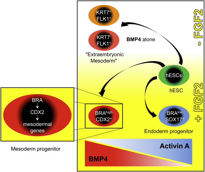
BMP is thought to induce hESC differentiation toward multiple lineages including mesoderm and trophoblast. The BMP-induced trophoblast phenotype is a long-standing paradox in stem cell biology. Here we readdressed BMP function in hESCs and mouse epiblast-derived cells. We found that BMP4 cooperates with FGF2 (via ERK) to induce mesoderm and to inhibit endoderm differentiation. These conditions induced cells with high levels of BRACHYURY (BRA) that coexpressed CDX2. BRA was necessary for and preceded CDX2 expression; both genes were essential for expression not only of mesodermal genes but also of trophoblast-associated genes. Maximal expression of the latter was seen in the absence of FGF but these cells coexpressed mesodermal genes and moreover they differed in cell surface and epigenetic properties from placental trophoblast. We conclude that BMP induces human and mouse pluripotent stem cells primarily to form mesoderm, rather than trophoblast, acting through BRA and CDX2.


Figures

None
Graphical abstract
Figure 1
Figure 1
BMP and FGF Cooperate to Induce BRA and CDX2 (A) qPCR analysis of hESCs grown as indicated. Colored bars represent the absence (blue) or presence (red) of BMP4. p ≤ 0.05; +p ≤ 0.01; t test. (B) Immunoblots of hESCs grown as indicated, where “Sb” refers to inhibition of Activin/Nodal signaling by SB431542, “-” is no Activin addition and “10” or “100” is the ng/ml Activin. (C) qPCR analysis (i) and immunoblots (ii) of hESCs grown as indicated; “Uo” is ERK inhibitor UO126. In (i) p ≤ 0.05; +p ≤ 0.01; t test; comparisons done to day 0 are indicated in black, and comparisons between the two indicated treatments are in red. (D) qPCR analysis of hESCs grown as indicated; pluripotency conditions (day 0, D0), CDM alone, DMSO control, FLyB, or FLyA plus other factors as indicated below each histogram (Nog, Noggin at 200 ng/ml; B, BMP at 10, 50, or 100 ng/ml; Sb, SB431542 at 10 μM; A, Activin at 10, 50, or 100 ng/ml). p ≤ 0.05; +p ≤ 0.01; t test. (E) Representative fluorescent images of hESCs grown as indicated. Samples were immunostained for BRA, FOXA2, and CDX2. See also Figure S1.
Figure 2
Figure 2
Distinct BRAhigh/CDX2+ and BRAlow/SOX17+ Populations Emerge from FLyA and FLyB Treatments (A) Flow cytometry histograms of hESCs grown as indicated. Circles represent subpopulations of BRAhigh and BRAlow cells, which colocalize with CDX2 or SOX17, respectively. (B) Microarray gene expression heat map of undifferentiated (day 0, D0) versus differentiated hESCs grown as indicated. Heat-map colors indicate log2 fold-changes. (C) Flow cytometry histograms showing BRA coexpression with HAND1, MESP1, or FOXA2 in hESCs grown as indicated. (Di) qPCR analysis of hESCs induced to differentiate toward proximal streak mesoderm (D5) and untreated controls (pluripotency conditions - D0). (Dii) Representative fluorescent image of D5 hESCs grown as indicated. Samples were immunostained for CDX2. (E) qPCR analysis of hESCs differentiated as indicated. p ≤ 0.05; +p ≤ 0.01; t test. Comparisons were done between the two indicated treatments. (F) qPCR analysis of hESCs differentiated as indicated. See also Figure S2.
Figure 3
Figure 3
Expression of Both Embryonic and Extraembryonic Lineage-Associated Genes in BMP-Treated hESCs Depends on a BRA-Driven Gene Regulatory Network (A) qPCR analysis of sh-BRA knockdown (BRA KD) hESC lines and a scrambled control line (Scramble) grown as indicated. p ≤ 0.05; +p ≤ 0.01; t test. (B) Representative fluorescent images of BRA KD and control hESCs grown as indicated. Samples were immunostained for CDX2, BRA, and DAPI. (C) qPCR analysis of sh-BRA KD and control hESCs grown as indicated to induce expression of extraembryonic genes. p ≤ 0.05; +p ≤ 0.01; t test. (D) qPCR analysis of sh-CDX2 knockdown (CDX2 KD) hESC lines and a control line grown as indicated. p ≤ 0.05; +p ≤ 0.01; t test. (E) Representative fluorescent images of CDX2 KD and control hESCs grown as indicated. Samples were immunostained for CDX2, BRA, and DAPI. (F) qPCR analysis of CDX2 KD and control hESCs differentiated as indicated. p ≤ 0.05; +p ≤ 0.01; t test. (G) qPCR analysis of CDX2 KD and control hESCs grown as indicated. p ≤ 0.05; +p ≤ 0.01; t test. See also Figure S3.
Figure 4
Figure 4
Characteristics of BMP-Treated hESCs Distinguish Them from Placental Trophoblast (Ai) qPCR analysis of ELF5 expression in hESCs grown as indicated. (Aii) Representative fluorescent images of hESCs grown as indicated. Samples were immunostained for ELF5. (B) Bisulphite sequencing analysis of the ELF5 promoter region in hESCs grown as indicated. Filled circles indicate methylated cytosine residues. Top row: hypomethylation status in placental trophoblast. Distance from ATG is shown in base pairs. (C) Table of HLA class I gene expression in placental (in vivo) trophoblast (Apps et al., 2009) and hESCs grown in pluripotency (day 0, D0) or in differentiating conditions as indicated. Legend: -, no expression (0%–4%); +/−, 5%–30%; +, 31%–50%; ++, 51%–70%; +++, 71%–100% of the cells, as determined by flow cytometry. (D) Representative images of placental serial sections stained for HLA-G or the 1B10-fibroblast antigen (1B10-Fib). Arrows indicate HLA-G-positive extravillous trophoblast cells; arrowheads indicate 1B10-fibroblast-negative villous trophoblast. (E) Flow cytometry histograms showing class I HLA expression (detected by W6/32 antibody) and the epitope detected by 1B10-fibroblast antibody (1B10-Fib) in hESCs grown in pluripotency conditions (hESCs) or in differentiating conditions as indicated. See also Figure S4.
Figure 5
Figure 5
Cells Expressing HCGα, GCM1, and KRT7 Represent a Subpopulation of Mesoderm Cells (A) qPCR analysis of KRT7+ and KRT7 cells sorted from hESCs grown as indicated. (B) Representative fluorescent images of hESCs grown as indicated. Samples were immunostained for KRT7, ISL1, and the nuclear marker DAPI. (C) Flow cytometry histograms showing FLK1 (upper panels), VCAM-1 (lower panels), and KRT7 coexpression in hESCs grown as indicated. (D) qPCR analysis of mouse late epiblast explants grown in pluripotency (A, ActivinA, 10 ng/ml; F, FGF2, 20 ng/ml) or differentiation conditions as indicated. p ≤ 0.05; +p ≤ 0.01; t test. (Ei) Representative fluorescent image of human placental mesenchyme plated for a week in serum-containing medium. Samples were immunostained for KRT7 and with DAPI. (Eii) qPCR analysis of human placental mesenchyme before plating cells to generate outgrowths. (F) Representative light and fluorescent images of hESCs differentiated as indicated. White arrowheads point to multinucleated cells. (G) Flow cytometry histograms showing KRT7 expression, class 1 HLA epitopes (W6/32 antibody), class I A and B HLA epitopes (Tu155 antibody), and the epitope detected by 1B10-fibroblast (1B10-Fib) in hESCs grown as indicated. See also Figure S5.

Comment in

  • When BMP meets FGF.
    Greber B. Greber B. Cell Stem Cell. 2011 Aug 5;9(2):91-2. doi: 10.1016/j.stem.2011.07.004. Cell Stem Cell. 2011. PMID: 21816358

References

    1. Apps R., Murphy S.P., Fernando R., Gardner L., Ahad T., Moffett A. Human leucocyte antigen (HLA) expression of primary trophoblast cells and placental cell lines, determined using single antigen beads to characterize allotype specificities of anti-HLA antibodies. Immunology. 2009;127:26–39. - PMC - PubMed
    1. Apps R., Sharkey A., Gardner L., Male V., Trotter M., Miller N., North R., Founds S., Moffett A. Genome-wide expression profile of first trimester villous and extravillous human trophoblast cells. Placenta. 2011;32:33–43. - PMC - PubMed
    1. Beck F., Erler T., Russell A., James R. Expression of Cdx-2 in the mouse embryo and placenta: possible role in patterning of the extra-embryonic membranes. Dev. Dyn. 1995;204:219–227. - PubMed
    1. Beddington R.S., Robertson E.J. An assessment of the developmental potential of embryonic stem cells in the midgestation mouse embryo. Development. 1989;105:733–737. - PubMed
    1. Beddington R.S., Rashbass P., Wilson V. Brachyury—a gene affecting mouse gastrulation and early organogenesis. Dev. Suppl. 1992:157–165. - PubMed

Publication types

MeSH terms