. 2011 Oct 1;71(19):6184-94.
doi: 10.1158/0008-5472.CAN-11-1341. Epub 2011 Aug 4.

Wnt5a suppresses epithelial ovarian cancer by promoting cellular senescence

Affiliations

Wnt5a suppresses epithelial ovarian cancer by promoting cellular senescence

Benjamin G Bitler et al. Cancer Res. .

Abstract

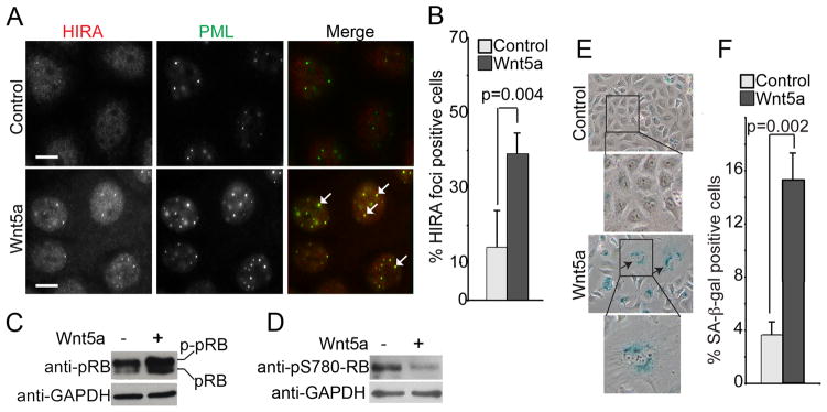
Epithelial ovarian cancer (EOC) remains the most lethal gynecologic malignancy in the United States. Thus, there is an urgent need to develop novel therapeutics for this disease. Cellular senescence is an important tumor suppression mechanism that has recently been suggested as a novel mechanism to target for developing cancer therapeutics. Wnt5a is a noncanonical Wnt ligand that plays a context-dependent role in human cancers. Here, we investigate the role of Wnt5a in regulating senescence of EOC cells. We show that Wnt5a is expressed at significantly lower levels in human EOC cell lines and in primary human EOCs (n = 130) compared with either normal ovarian surface epithelium (n = 31; P = 0.039) or fallopian tube epithelium (n = 28; P < 0.001). Notably, a lower level of Wnt5a expression correlates with tumor stage (P = 0.003) and predicts shorter overall survival in EOC patients (P = 0.003). Significantly, restoration of Wnt5a expression inhibits the proliferation of human EOC cells both in vitro and in vivo in an orthotopic EOC mouse model. Mechanistically, Wnt5a antagonizes canonical Wnt/β-catenin signaling and induces cellular senescence by activating the histone repressor A/promyelocytic leukemia senescence pathway. In summary, we show that loss of Wnt5a predicts poor outcome in EOC patients and Wnt5a suppresses the growth of EOC cells by triggering cellular senescence. We suggest that strategies to drive senescence in EOC cells by reconstituting Wnt5a signaling may offer an effective new strategy for EOC therapy.


Figures

Figure 1
Figure 1. Wnt5a is expressed at significantly lower levels in human EOC cells compared with normal human ovarian surface or fallopian tube epithelial cells, and a lower level of Wnt5a expression predicts shorter overall survival in human EOC patients
(A) Expression of Wnt5a mRNA in primary human ovarian surface epithelial (HOSE) cells and the indicated human EOC cell lines was determined by semi-quantitative RT-PCR. Expression of GAPDH mRNA was used a loading control. (B) Wnt5a mRNA levels were quantified by quantitative RT-PCR in six individual isolations of primary HOSE cells and seven different EOC cell lines. Expression of β-2-microglobulin was used to normalize Wnt5a mRNA expression. * p = 0.008 compared with human EOC cells. (C) Same as (A), but examined for Wnt5a and GAPDH protein expression by immunoblotting. (D) Examples of Wnt5a IHC staining in normal human ovarian surface epithelium, fallopian tube epithelium, and EOC of indicated histological subtypes. Bar = 50 μm. Arrows point to examples of positively stained human ovarian surface epithelial cells and fallopian tube epithelial cells. (E) Representative images from tissue microarray depicting low Wnt5a expression correlated with high Ki67, a cell proliferation marker. Bar = 50 μm. (F) Loss of Wnt5a expression is an independent poor prognosis marker in human EOC patients. A lower level of Wnt5a expression correlates with shorter overall survival in human EOC patients. The univariate overall survival curve (Kaplan-Meier method) for EOC patients (n = 123) with high or low Wnt5a expression as determined by immunohistochemical analysis.
Figure 2
Figure 2. Promoter DNA CpG island hypermethylation contributes to Wnt5a downregulation in human EOC cells
(A) Schematic structure of the human Wnt5a gene transcript and its promoter CpG islands. Locations of exon 1 (open rectangle), CpG sites (vertical lines) and coding exons (filled rectangle) and the transcription start site (curved arrow) are indicated. Flat arrows indicated the positions of primers used for PCR amplification, and the size of PCR product is also indicated. (B) PEO1 cells were treated with 5 μM AzaC for 4 days, and mRNA was isolated from control and AzaC treated cells and examined for Wnt5a mRNA expression by qRT-PCR. Mean of three independent experiments with SD. (C) Same as (C) but examined for Wnt5a protein expression by immunoblotting.
Figure 3
Figure 3. Wnt5a restoration inhibits the growth of human EOC cells by antagonizing canonical Wnt/β-catenin signaling
(A) OVCAR5 cells were transduced with a control or Wnt5a encoding puromycin resistant retrovirus. The infected cells were drug-selected with 3 μg/ml puromycin. Expression of Wnt5a mRNA in drug-selected cells was determined by qRT-PCR. (B) Same as (A), but examined for expression of Wnt5a and β-actin in control or Wnt5a-infected OVCAR5 and primary HOSE cells by immunoblotting. Relative levels of Wnt5a expression was indicated based on densometric analysis using NIH Image J software. (C) Same as (A), but equal number (5000) of drug-selected control (open triangles and solid line) or Wnt5a-infected cells (open circles and dotted line) were cultured on plastic plates for 4 days, and the number of cells was counted (Control ± SD or Wnt5a ± SD (n=3); student t-test was used for calculating p value) at day 1 (6,666±1,258 vs. 5,000±1,000; p = 0.1469), day 2 (14,583±954 vs. 9,583±3,463; p = 0.084), day 3 (41,250±6,538 vs. 14,750±2,787; *p = 0.0038) and day 4 (83,055±8,978 vs. 35,416±2,055; **p = 0.001). Mean of three independent experiments with SD and linear regression. (D) Same as (C), but grown under anchorage-independent condition in soft-agar. The number of colonies was counted 2 weeks after initial inoculation. Mean of three independent experiments with SD. (E) Same as (A), but examined for the levels of soluble β-catenin and β-actin expression by immunoblotting (NT = non-treated). (F) Same as (A), but examined for expression of indicated β-catenin target genes by qRT-PCR. Expression of β-2- microglobulin was used to normalize the expression of indicated genes. * p = 0.0095, ** p = 0.0012 and *** p = 0.0286 compared with controls.
Figure 4
Figure 4. Wnt5a promotes senescence of primary HOSE cells
(A) Young proliferating primary HOSE cells were passaged to senescence (after seven population doublings). Expression of SA-β-gal activity was measured in young and naturally senescent primary HOSE cells. (B) Same as (A). Quantitation of SA-β-gal positive cells (**p < 0.001). (C) Same as (A), but mRNA was isolated and examined for Wnt5a expression by qRT-PCR. Expression of β-2-microglobulin (B2M) was used as a control. (*p = 0.003). (D) Young primary HOSE cells were transduced with retrovirus encoding human Wnt5a gene or a control. Expression of Wnt5a in indicated cells was determined by qRT-PCR. Expression of β-2-microglobulin (B2M) was used as a control. (E) Same as (D), but stained for expression of SA-β-gal activity in drug-selected cells. (F) Quantitation of (E). Mean of three independent experiments with SD. * p<0.05.
Figure 5
Figure 5. Wnt5a restoration triggers cellular senescence in human EOC cells
(A) Control and Wnt5a expressing OVCAR5 EOC cells were stained with antibodies to HIRA and PML. Arrows point to examples of co-localized HIRA and PML bodies. Bar = 10 μm. (B) Quantitation of (A). 200 cells from control and Wnt5a expressing cells were examined for HIRA and PML co-localization. Mean of three independent experiments with SD. (C) Same as (A), but examined for pRB and GAPDH expression. (D) Same as (C), but examined for pRBpS780 and GAPDH expression. (E) Same as (A), but examined for senescence-associated β-galactosidase (SA-β-gal) activity. (F) Quantitation of (E). Mean of three independent experiments with SD.
Figure 6
Figure 6. Wnt5a restoration inhibits tumor growth and promotes senescence of human EOC cells in vivo
(A) OVCAR5 cells were transduced with luciferase encoding hygromycin resistant retrovirus together with a control or Wnt5a encoding puromycin-resistant retrovirus. Drug-selected cells were unilaterally injected into the peri-ovarian bursa sac of the female immuno-compromised mice (n = 6 for each of the groups). The radiance of luciferase bioluminescence, an indicator of the rate for tumor growth, was measured every 5 day from day 10 until day 30 using the IVIS imaging system. Shown are images taken at day 10 and day 30, respectively. (B) Quantitation of tumor growth from injected OVCAR5 cells expressing Wnt5a or control at indicated time points. * p = 0.038 compared with controls. (C) Following tumor dissection, expression of Wnt5a in tumors formed by control or Wnt5a expressing OVCAR5 EOC cells was determined by immunohistochemical staining against Wnt5a (40X). Bar = 50 μm. (D) Same as (C), but examined for expression of Ki67, a marker of cell proliferation (40X). Bar = 50 μm. (E) Quantitation of (D). * p = 0.008 compared with controls. (F) Expression of SA-β-gal activity was examined on sections of fresh-frozen tumors formed by OVCAR5 cells expressing control or Wnt5a (40X). Bar = 100 μm. (G) Quantitation of (F). * p = 0.003 compared with controls.

Similar articles

Cited by

References

    1. Kuilman T, Michaloglou C, Mooi WJ, Peeper DS. The essence of senescence. Genes Dev. 2010;24:2463–79. - PMC - PubMed
    1. Adams PD. Healing and hurting: molecular mechanisms, functions, and pathologies of cellular senescence. Mol Cell. 2009;36:2–14. - PubMed
    1. Dimri GP, Lee X, Basile G, Acosta M, Scott G, Roskelley C, et al. A biomarker that identifies senescent human cells in culture and in aging skin in vivo. Proc Natl Acad Sci U S A. 1995;92:9363–7. - PMC - PubMed
    1. Ventura A, Kirsch DG, McLaughlin ME, Tuveson DA, Grimm J, Lintault L, et al. Restoration of p53 function leads to tumour regression in vivo. Nature. 2007;445:661–5. - PubMed
    1. Xue W, Zender L, Miething C, Dickins RA, Hernando E, Krizhanovsky V, et al. Senescence and tumour clearance is triggered by p53 restoration in murine liver carcinomas. Nature. 2007;445:656–60. - PMC - PubMed

Publication types

MeSH terms