Review
. 2011 Aug 5;109(4):463-74.
doi: 10.1161/CIRCRESAHA.110.225003.

Status and prospects for discovery and verification of new biomarkers of cardiovascular disease by proteomics

Affiliations
Review

Status and prospects for discovery and verification of new biomarkers of cardiovascular disease by proteomics

Robert E Gerszten et al. Circ Res. .

Abstract

Despite unmet needs for cardiovascular biomarkers, few new protein markers have been approved by the US Food and Drug Administration for the diagnosis or screening of cardiovascular diseases. Mass spectrometry-based proteomics technologies are capable of identifying hundreds to thousands of proteins in cells, tissues, and biofluids. Proteomics may therefore provide the opportunity to elucidate new biomarkers and pathways without a prior known association with cardiovascular disease; however, important obstacles remain. In this review, we focus on emerging techniques that may form a coherently integrated pipeline to overcome present limitations to both the discovery and validation processes.


Conflict of interest statement

Disclosures: The authors have no conflicts of interest related to this manuscript.

Figures

Figure 1
Figure 1
Overview of “Discovery Proteomics” by differential protein expression profiling using liquid chromatography coupled to tandem mass spectrometry, LC-MS/MS (see text).
Figure 1
Figure 1
Overview of “Discovery Proteomics” by differential protein expression profiling using liquid chromatography coupled to tandem mass spectrometry, LC-MS/MS (see text).
Figure 2
Figure 2
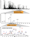
The LC-MS/MS data acquisition process commonly used in biomarker discovery. Top panel: illustrates the Total Ion Current (TIC) trace recorded during the on-line LC-MS/MS analysis of a tissue lysate. The TIC is similar to a UV trace from the chromatograph, but instead of a plot of absorbance vs time, the TIC is a plot of the sum of the ions entering the MS vs. time. Middle panel: mass spectrum (plot of m/z vs. time) recorded in ca. 1 s at time = 70.55 min during the LC-MS experiment. The peaks correspond to the m/z of peptides eluting from the column at this moment in time. On modern, high performance instruments the MS data are recorded at high resolution and with high mass precision, typically 30,000 – 60,000 resolution and 2–5 ppm (part-per-million) mass accuracy. Both high resolution and mass accuracy are important in the analysis of highly complex mixtures such as digested plasma. In such complex mixtures, many peptides of different amino acid sequence will have the same nominal molecular mass (i.e., within 1 mass unit) but will differ in their accurate molecular masses. High instrument resolving power enables peptides of similar but non-identical mass to be separated on the m/z scale, and high mass accuracy facilitates their identification by the software by narrowing the list of possible amino acid compositions that could correspond to the observed accurate mass. Bottom panel: one of eight MS/MS spectra recorded in ca. 100 ms by the on-board processors in the MS system. In a typical experiment, the top 8–10 most abundant ions in the current mass spectrum are sequentially mass selected and their fragmentation spectra recorded for sequencing automatically during a 1 to 3 second period. After acquisition of the current block of “n” MS/MS spectra, the scan cycle is repeated continuously for the duration of the on-line LC separation, each time starting with acquisition of a full scan MS spectrum followed by “n” more MS/MS spectra. These data are subsequently analyzed by the analysis software (e.g., Mascot, Spectrum Mill, Sequest, XTandem, etc.). The peptide sequence determined by the data analysis software is shown. The b- and y-ions correspond to fragmentation along the peptide backbone at amide bonds with charge retained on the N-terminal fragment or C-terminal fragment, respectively.
Figure 3
Figure 3
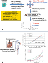
Panel A. An overview of the SISCAPA method (Stable Isotope Standards and Capture by Anti-Peptide Antibodies). Bead-bound anti-peptide antibodies made against selected tryptic peptides from protein biomarkers of interest are added to trypsin-digested plasma along with a known amounts of stable isotope-labeled versions of each analyte peptide. The antibodies capture and enrich the heavy-labeled exogenous form and sample-derived form of the peptides. After washing, the peptides are eluted from the beads into the MS system and the relative amounts of each analyte peptide is measured by multiple reaction monitoring mass spectrometry (MRM-MS). Panel B. In the upper right, the multiplex SISCAPA assay for peptides derived from Cardiac Troponin I (cTnI) and IL-33 is shown. Limits of quantification for both proteins are approximately 1 nanogram/mL in human plasma. The lower right panel shows concentrations of cTnI as determined by SISCAPA in patients undergoing a planned myocardial infarct (PMI). Six time points were sampled over the course of the procedure (0, 0.2h, 1h, 2h, 4h, and 24h). Each measurement is an average of three injections; error bars represent the standard deviation. A non-linear scale was used for the time axis.

Similar articles

Cited by

References

    1. Khot UN, Khot MB, Bajzer CT, Sapp SK, Ohman EM, Brener SJ, Ellis SG, Lincoff AM, Topol EJ. Prevalence of conventional risk factors in patients with coronary heart disease. JAMA. 2003;290:898–904. - PubMed
    1. Antman E, Grudzien C, Sacks DB. Evaluation of a rapid bedside assay for detection of serum cardiac troponin T. JAMA. 1995;273:1279–1282. - PubMed
    1. Lindahl B, Toss H, Siegbahn A, Venge P, Wallentin L for the FRISC Study Group. Fragmin during Instability in Coronary Artery Disease. Markers of myocardial damage and inflammation in relation to long-term mortality in unstable coronary artery disease. N Engl J Med. 2000;343:1139–1147. - PubMed
    1. Antman EM, Tanasijevic MJ, Thompson B, Schactman M, McCabe CH, Cannon CP, Fischer GA, Fung AY, Thompson C, Wybenga D, Braunwald E. Cardiac-specific troponin I levels to predict the risk of mortality in patients with acute coronary syndromes. N Engl J Med. 1996;335:1342–1349. - PubMed
    1. Hamm CW, Heeschen C, Goldmann B, Vahanian A, Adgey J, Miguel CM, Rutsch W, Berger J, Kootstra J, Simoons ML for the c7E3 Fab Antiplatelet Therapy in Unstable Refractory Angina (CAPTURE) Study Investigators. Benefit of abciximab in patients with refractory unstable angina in relation to serum troponin T levels. N. Engl. J. Med. 1999;340:1623–1629. - PubMed

Publication types