. 2011;6(7):e22615.
doi: 10.1371/journal.pone.0022615. Epub 2011 Jul 26.

Conformational plasticity of proNGF

Affiliations

Conformational plasticity of proNGF

Francesca Paoletti et al. PLoS One. 2011.

Abstract

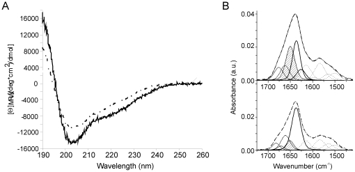
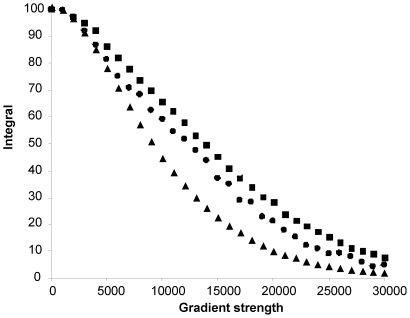
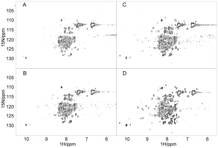
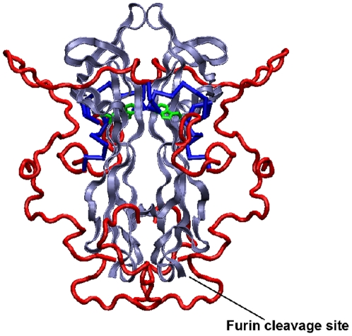
Nerve Growth Factor is an essential protein that supports neuronal survival during development and influences neuronal function throughout adulthood, both in the central and peripheral nervous system. The unprocessed precursor of NGF, proNGF, seems to be endowed with biological functions distinct from those of the mature protein, such as chaperone-like activities and apoptotic and/or neurotrophic properties. We have previously suggested, based on Small Angle X-ray Scattering data, that recombinant murine proNGF has features typical of an intrinsically unfolded protein. Using complementary biophysical techniques, we show here new evidence that clarifies and widens this hypothesis through a detailed comparison of the structural properties of NGF and proNGF. Our data provide direct information about the dynamic properties of the pro-peptide and indicate that proNGF assumes in solution a compact globular conformation. The N-terminal pro-peptide extension influences the chemical environment of the mature protein and protects the protein from proteolytic digestion. Accordingly, we observe that unfolding of proNGF involves a two-steps mechanism. The distinct structural properties of proNGF as compared to NGF agree with and rationalise a different functional role of the precursor.


Conflict of interest statement

Competing Interests: The authors have declared that no competing interests exist.

Figures

Figure 1
Figure 1. CD and FT-IR analysis of NGF and proNGF25.
A) Far-UV CD spectra of NGF (black line) and proNGF25 (grey line) in 50 mM sodium phosphate. The difference curve proNGF25 subtracted NGF is represented by the dashed curve. B) Curve fittings of FT-IR spectra of proNGF25 (upper panel) and NGF (lower panel) in deuterated phosphate buffer at pD 7. The component bands present in the amide I' region are assigned to β-sheet (bold lines), to random domains (dashed line) and to turns (dot-dashed lines). The component bands in the 1600–1500 cm-1 region are attributed to side-chain and amide II contributions and are shown for eye guideline only. The sum of all these components is represented as a dashed line.
Figure 2
Figure 2. NMR spectral properties of NGF and proNGF25.
A) Comparison of 1D spectra for NGF (lower spectrum) and proNGF25 (upper spectrum). B) Expansion of the high-field regions of the spectra shown in A) and comparison with the same regions of the spectra of ataxin-3 (35 kDa) and its N-terminal Josephin domain (20 kDa). The stacked plot shows from bottom up the spectra of NGF, proNGF25, Josephin and ataxin-3 respectively. C) 2D HSQC heteronuclear spectra of NGF (bottom), proNGF25 (middle) and overlay of the two (top) (NGF is shown in black, proNGF25 in red). The spectra were recorded on 0.4 mM protein concentrations, 25°C and at 800 MHz overnight.
Figure 3
Figure 3. NMR diffusion data.
The plot reports the integral of an envelope of signals (y-axis) against the applied gradient strength (x-axis). The symbols correspond to the IscS dimer (grey squares), proNGF25 (black squares), DHFR (grey diamonds), NGF (black circles) and the NusA KH domain (black triangles). For each protein, the peak integrals were plot against the applied gradient strength.
Figure 4
Figure 4. Enzymatic cleavage of the pro-peptide as followed by NMR.
A) 2D HSQC heteronuclear spectrum of proNGF25 before addition of furin, B) immediately after furin addition, C) at completion of the cleavage reaction and D) after further addition of trypsin. The spectra were recorded at 25°C and 600 MHz. The initial sample was 0.1 mM, double labelled and less monodispersed than the one shown in Figure 2C (top and middle panels).
Figure 5
Figure 5. DSC thermograms of proNGF25 and NGF.
DSC thermograms were recorded for solutions in a saline phosphate buffer at pH 7. A) NGF at 20 µM and heating rates of 90°C/h (bold line) and 60°C/h (thin line), B) proNGF25 at 20 µM and heating rates 90°C/h (bold line) and 60°C/h (thin line).
Figure 6
Figure 6. Model of proNGF25.
The model was built according to previous SAXS data . Mature NGF is shown in blue, the pro-peptide domain in red. The side chain of NGF Trp21 is shown in green. This residue was suggested to take part to the interaction of the NGF moiety with the pro-peptide . The position of the furin cleavage site is also indicated.

References

    1. Levi-Montalcini R. The nerve growth factor 35 years later. Science. 1987;237:1154–1162. - PubMed
    1. Teng KK, Hempstead BL. Neurotrophins and their receptors: signaling trios in complex biological systems. Cell Mol Life Sci. 2004;61:35–48. - PMC - PubMed
    1. Chao MV, Bothwell MA, Ross AH, Koprowski H, Lanahan AA, et al. Gene transfer and molecular cloning of the human NGF receptor. Science. 1986;232:518–521. - PubMed
    1. He XL, Garcia KC. Structure of nerve growth factor complexed with the shared neurotrophin receptor p75. Science. 2004;304:870–875. - PubMed
    1. Wiesmann C, Ultsch MH, Bass SH, de Vos AM. Crystal structure of nerve growth factor in complex with the ligand-binding domain of the TrkA receptor. Nature. 1999;401:184–188. - PubMed

Publication types

MeSH terms