

Synaptic responses evoked by tactile stimuli in Purkinje cells in mouse cerebellar cortex Crus II in vivo
- PMID: 21818384
- PMCID: PMC3144243
- DOI: 10.1371/journal.pone.0022752
Synaptic responses evoked by tactile stimuli in Purkinje cells in mouse cerebellar cortex Crus II in vivo
Abstract
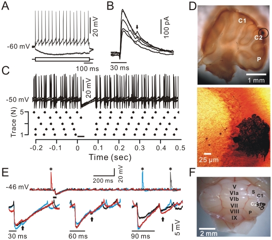
Background: Sensory stimuli evoke responses in cerebellar Purkinje cells (PCs) via the mossy fiber-granule cell pathway. However, the properties of synaptic responses evoked by tactile stimulation in cerebellar PCs are unknown. The present study investigated the synaptic responses of PCs in response to an air-puff stimulation on the ipsilateral whisker pad in urethane-anesthetized mice.
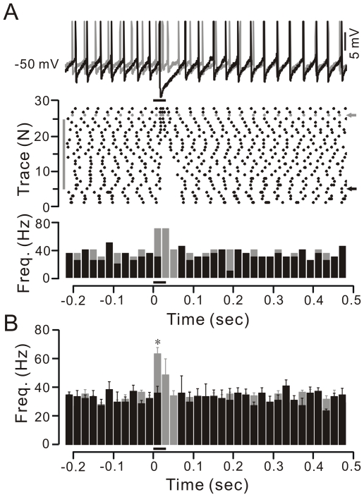
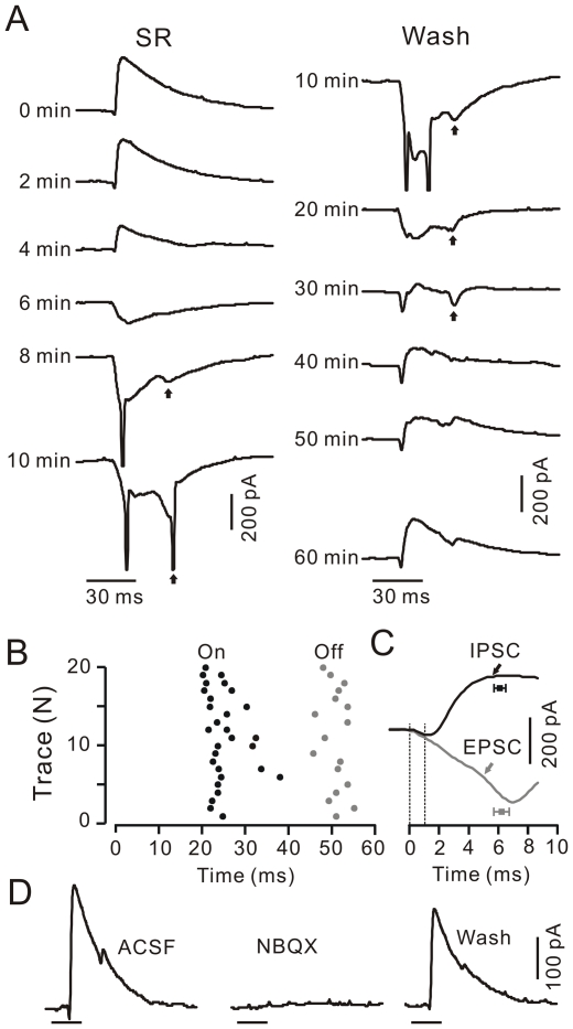
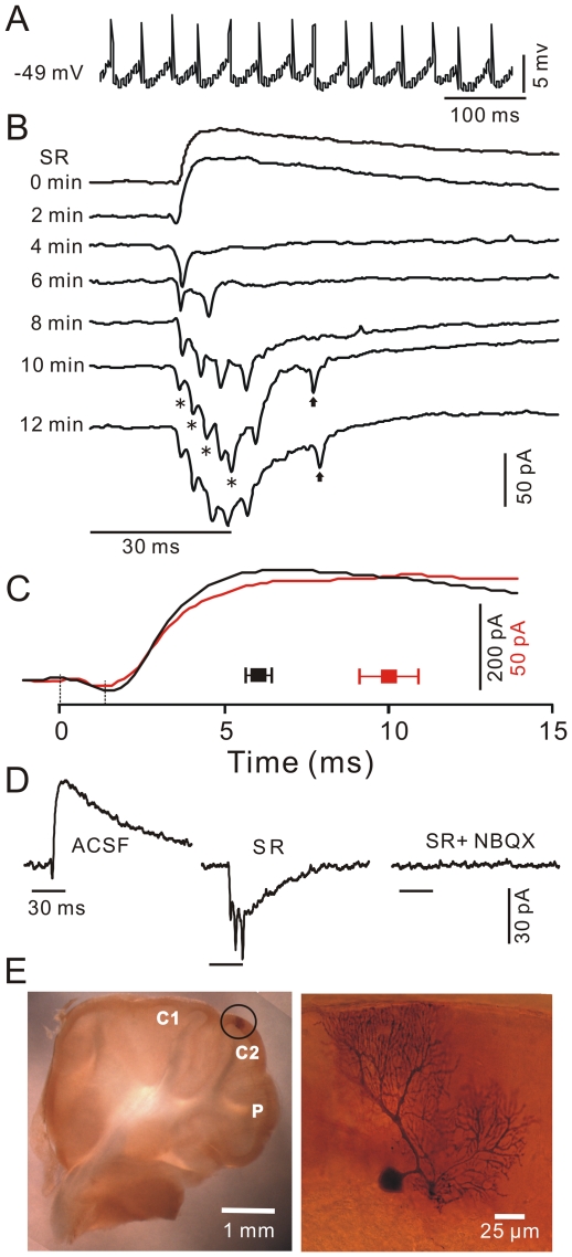
Methods and main results: Thirty-three PCs were recorded from 48 urethane-anesthetized adult (6-8-week-old) HA/ICR mice by somatic or dendritic patch-clamp recording and pharmacological methods. Tactile stimulation to the ipsilateral whisker pad was delivered by an air-puff through a 12-gauge stainless steel tube connected with a pressurized injection system. Under current-clamp conditions (I = 0), the air-puff stimulation evoked strong inhibitory postsynaptic potentials (IPSPs) in the somata of PCs. Application of SR95531, a specific GABA(A) receptor antagonist, blocked IPSPs and revealed stimulation-evoked simple spike firing. Under voltage-clamp conditions, tactile stimulation evoked a sequence of transient inward currents followed by strong outward currents in the somata and dendrites in PCs. Application of SR95531 blocked outward currents and revealed excitatory postsynaptic currents (EPSCs) in somata and a temporal summation of parallel fiber EPSCs in PC dendrites. We also demonstrated that PCs respond to both the onset and offset of the air-puff stimulation.
Conclusions: These findings indicated that tactile stimulation induced asynchronous parallel fiber excitatory inputs onto the dendrites of PCs, and failed to evoke strong EPSCs and spike firing in PCs, but induced the rapid activation of strong GABA(A) receptor-mediated inhibitory postsynaptic currents in the somata and dendrites of PCs in the cerebellar cortex Crus II in urethane-anesthetized mice.
Conflict of interest statement
Figures
Similar articles
-
Roles of molecular layer interneurons in sensory information processing in mouse cerebellar cortex Crus II in vivo.PLoS One. 2012;7(5):e37031. doi: 10.1371/journal.pone.0037031. Epub 2012 May 18. PLoS One. 2012. PMID: 22623975 Free PMC article.
-
Sensory stimulus evokes inhibition rather than excitation in cerebellar Purkinje cells in vivo in mice.Neurosci Lett. 2011 Jan 7;487(2):182-6. doi: 10.1016/j.neulet.2010.10.018. Epub 2010 Oct 19. Neurosci Lett. 2011. PMID: 20965231
-
Activation of α 2A and α 2B -adrenergic receptors inhibits tactile stimulation-evoked parallel fiber-Purkinje cell synaptic transmission in mouse cerebellar cortex.Neuroreport. 2024 Feb 7;35(2):115-122. doi: 10.1097/WNR.0000000000001983. Epub 2023 Dec 12. Neuroreport. 2024. PMID: 38109417 Free PMC article.
-
Pharmacology of the metabotropic glutamate receptor mediated current at the climbing fiber to Purkinje cell synapse.Prog Brain Res. 2005;148:299-306. doi: 10.1016/S0079-6123(04)48023-6. Prog Brain Res. 2005. PMID: 15661198 Review.
-
[Role of glutamate transporters in excitatory synapses in cerebellar Purkinje cells].Brain Nerve. 2007 Jul;59(7):669-76. Brain Nerve. 2007. PMID: 17663137 Review. Japanese.
Cited by
-
Fentanyl Inhibits Air Puff-Evoked Sensory Information Processing in Mouse Cerebellar Neurons Recorded in vivo.Front Syst Neurosci. 2020 Aug 4;14:51. doi: 10.3389/fnsys.2020.00051. eCollection 2020. Front Syst Neurosci. 2020. PMID: 32848643 Free PMC article.
-
Simple and complex spike responses of mouse cerebellar Purkinje neurons to regular trains and omissions of somatosensory stimuli.J Neurophysiol. 2021 Sep 1;126(3):763-776. doi: 10.1152/jn.00170.2021. Epub 2021 Aug 4. J Neurophysiol. 2021. PMID: 34346760 Free PMC article.
-
Roles of molecular layer interneurons in sensory information processing in mouse cerebellar cortex Crus II in vivo.PLoS One. 2012;7(5):e37031. doi: 10.1371/journal.pone.0037031. Epub 2012 May 18. PLoS One. 2012. PMID: 22623975 Free PMC article.
-
The indirect corticopontine pathway relays perioral sensory signals to the cerebellum via the mesodiencephalic junction.iScience. 2023 Jul 11;26(8):107301. doi: 10.1016/j.isci.2023.107301. eCollection 2023 Aug 18. iScience. 2023. PMID: 37539042 Free PMC article.
-
Facial Stimulation Induces Long-Term Potentiation of Mossy Fiber-Granule Cell Synaptic Transmission via GluN2A-Containing N-Methyl-D-Aspartate Receptor/Nitric Oxide Cascade in the Mouse Cerebellum.Front Cell Neurosci. 2022 Mar 30;16:863342. doi: 10.3389/fncel.2022.863342. eCollection 2022. Front Cell Neurosci. 2022. PMID: 35431815 Free PMC article.
References
-
- Bower JM, Woolston DC. Congruence of spatial organization of tactile projections to granule cell and Purkinje cell layers of cerebellar hemispheres of the albino rat: vertical organization of cerebellar cortex. J Neurophysiol. 1983;49:745–766. - PubMed
-
- Cook JR, Wiesendanger HC. Input from trigeminal cutaneous afferents to neurones of the inferior olive in rats. Exp Brain Res. 1976;26:193–202. - PubMed
-
- Eccles JC, Faber DS, Murphy JT, Sabah NH, Taborikova H. Investigations on integration of mossy fiber inputs to Purkyne cells in the anterior lobe. Exp Brain Res. 1971;13:54–77. - PubMed
-
- Palay SL, Chan-Palay V. New York: Springer-Verlag; 1974. Cerebellar Cortex.
Publication types
MeSH terms
LinkOut - more resources
Full Text Sources
