Review
. 2012 Jan 21;41(2):608-21.
doi: 10.1039/c1cs15112f. Epub 2011 Aug 4.

Misfolded proteins in Alzheimer's disease and type II diabetes

Affiliations
Review

Misfolded proteins in Alzheimer's disease and type II diabetes

Alaina S DeToma et al. Chem Soc Rev. .

Abstract

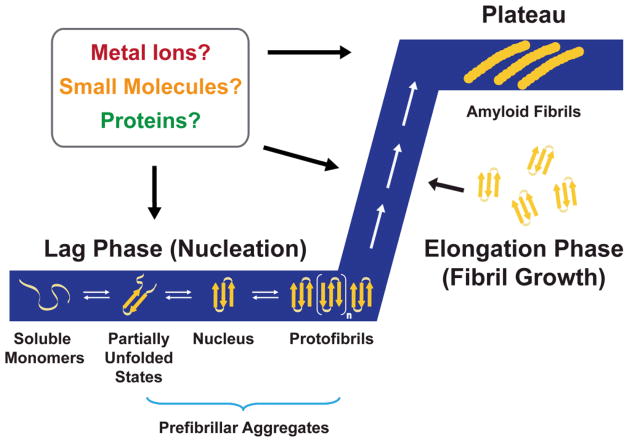
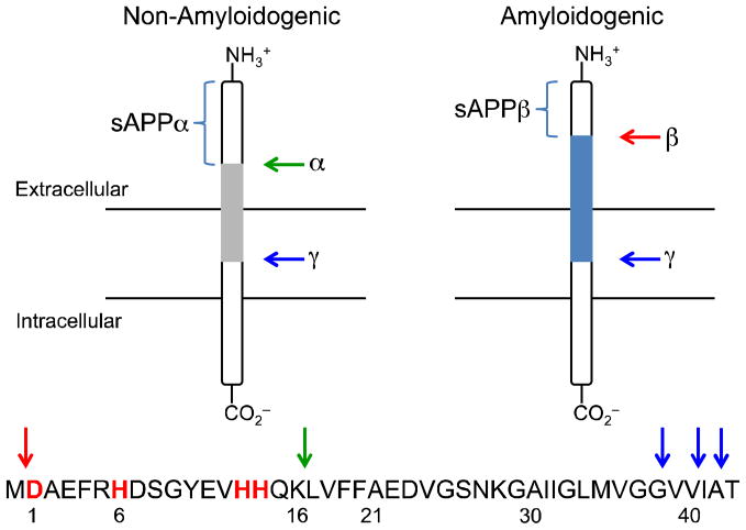
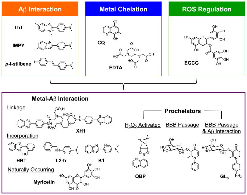
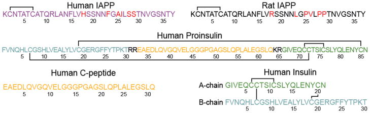
This tutorial review presents descriptions of two amyloidogenic proteins, amyloid-β (Aβ) peptides and islet amyloid polypeptide (IAPP), whose misfolding propensities are implicated in Alzheimer's disease (AD) and type II diabetes, respectively. Protein misfolding diseases share similarities, as well as some unique protein-specific traits, that could contribute to the initiation and/or development of their associated conditions. Aβ and IAPP are representative amyloidoses and are used to highlight some of the primary considerations for studying misfolded proteins associated with human diseases in this review. Among these factors, their physiological formation, aggregation, interactions with metal ions and other protein partners, and toxicity are presented. Small molecules that target and modulate the metal-Aβ interaction and neurotoxicity are included to illustrate one of the current approaches for uncovering the complexities of protein misfolding at the molecular level.


Figures

Fig. 1
Fig. 1
Nucleation polymerization model of amyloid aggregation. Soluble monomeric forms of the peptides, such as amyloid-β (Aβ) and islet amyloid polypeptide (IAPP), self-associate during the lag phase (nucleation). This forms a nucleus that can be rapidly extended during the fibril growth period called the elongation phase. Amyloid fibrils are the end product of this process, and their thermodynamic stability allows them to reach a stage called the plateau. The aggregation processes produce a sigmoidal kinetic trace that is depicted in the figure (blue). Additional factors, such as metal ions, small molecules, and other proteins, can be involved at several points during this process and may either facilitate or inhibit this process (see text). This figure is adapted from ref. [reproduced by permission of the Royal Society of Chemistry].
Fig. 2
Fig. 2
Cleavage of APP by α-,β-, and γ-secretases in the non-amyloidogenic and amyloidogenic pathways, respectively. The common full-length peptides, Aβ1-40 and Aβ1-42, in AD are formed through the amyloidogenic pathway.
Fig. 3
Fig. 3
ROS generation facilitated by redox cycling of Cu-bound Aβ under reducing conditions (Fenton chemistry).
Fig. 4
Fig. 4
Multifunctional small molecules that can be used to investigate Cu(II)/Zn(II)-associated Aβ species in AD by combining properties of Aβ interaction (orange), metal chelation (blue), and/or ROS regulation (green). Other structural moieties can be implemented in the design to introduce different reactivity (e.g., ROS-activated metal chelation ability (prochelators), enhanced BBB permeability). Multifunctional molecules are shown in the purple box. Flavonoids such as EGCG have demonstrated antioxidative properties. Abbreviations for compounds: ThT = thioflavin-T, 2-[4-(dimethylamino)phenyl]-3,6-dimethylbenzothiazolium; IMPY = 6-iodo-2-(4'-dimethylamino)phenylimidazo[1,2-a]pyridine; p-I-stilbene = N,N-dimethyl-4-[(1E)-2-(4-iodophenyl)ethenyl]benzenamine; CQ = 5-chloro-7-iodo-8-hydroxyquinoline; EDTA = N,N'-1,2-ethanediylbis[N-(carboxymethyl)]glycine; EGCG = epigallocatechin-3-gallate, XH1 = N,N-bis[2-[[2-[[4-(2-benzothiazolyl)phenyl]amino]-2-oxoethyl](carboxymethyl)amino]ethyl]glycine; HBT = 2-(2-hydroxyphenyl)benzothiazole; L2-b = N1,N1-dimethyl-N4-(pyridin-2-ylmethyl)benzene-1,4-diamine; K1 = 6-(imidazo-[1,2-a]pyridin- 2-yl)-N,N-dimethylpyridin-3-amine; QBP = quinoline boronic acid, pinanediol ester; the prochelator for BBB passage = 3-(D-glucopyranosyloxy)-2-methyl-1-phenyl-4(1H)-pyridinone; GL3 = 1-(4-aminophenyl)-3-(β-D-glucopyranosyloxy)-2-methyl-4(1H)-pyridinone.
Fig. 5
Fig. 5
Amino acid sequences of IAPP (human and rat), human proinsulin, human C-peptide, and human insulin. Solid lines represent disulfide bonds and residues highlighted in red differ between human and rat IAPP. C-peptide and insulin are cleaved from the same proinsulin fragment, as color-coded, with 4 proinsulin residues removed altogether during processing. Residues highlighted in color in this figure correspond to those in Figs. 7 and 8.
Fig. 6
Fig. 6
Concentration dependent effect of zinc on hIAPP1-37 aggregation kinetics and description of the two associated binding sites. Elongation rates, lag time, and relative amyloid formation were obtained by fitting ThT curves to a sigmoidal function. Red arrows correspond to processes that accelerate amyloid formation, blue arrows correspond to processes that inhibit amyloid formation, and dashes signify little change in aggregation kinetics upon addition of Zn(II). Length of arrows corresponds to the magnitude of the effect.
Fig. 7
Fig. 7
Structures of monomeric hIAPP1-37 in helix promoting solvent (A) and hIAPP1-29–MBP (maltose binding protein) fusion protein dimer (B) (PDB 3G7V). The addition of Zn(II) to monomeric hIAPP in the helical conformation (A) induces a kink near the H18 Zn(II) binding site, resulting in a helix-kink-helix motif that is similar to the helix-kink-helix motif observed in the hIAPP moiety of the fusion protein dimer (magenta in B). The H18 side chain of hIAPP is displayed for reference and disulfide bonds are shown in yellow.
Fig. 8
Fig. 8
Structures of monomeric human insulin (A) (PDB 2JV1), monomeric proinsulin (B) (PDB 2KQP), and human insulin in the hexameric T6 conformation (C and D) (PDB 1MSO) with similar color schemes. Monomeric insulin is depicted with the A-chain in green, the B-chain in cyan/blue, disulfide bonds in yellow, and the region exhibiting the largest chemical shift perturbations upon binding to rIAPP (residues 10–20) are in blue. Side chains with the greatest chemical shift perturbations are shown as well. The presence of additional residues in proinsulin (orange in B) have little effect on the structure of the insulin moiety in proinsulin (cyan and green). The hypothesized interaction interface between monomeric insulin and IAPP would be located in the interior of the hexameric T6 insulin storage structure (C and D) and would be inaccessible to IAPP (dark blue with E13 side chain indicated for reference).

References

    1. Soto C. FEBS Lett. 2001;498:204–207. - PubMed
    1. Soto C. Nat Rev Neurosci. 2003;4:49–60. - PubMed
    1. Ross CA, Poirier MA. Nat Med. 2004;10:S10–S17. - PubMed
    1. Chiti F, Dobson CM. Annu Rev Biochem. 2006;75:333–366. - PubMed
    1. Fändrich M. Cell Mol Life Sci. 2007;64:2066–2078. - PMC - PubMed

Publication types

MeSH terms