

Spontaneous nonthymic tumors in SCID mice
- PMID: 21819692
- PMCID: PMC3123755
Spontaneous nonthymic tumors in SCID mice
Abstract
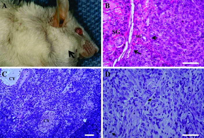
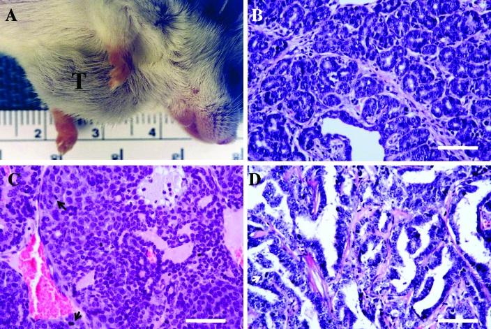
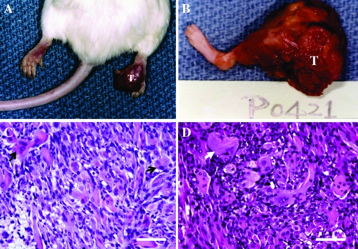
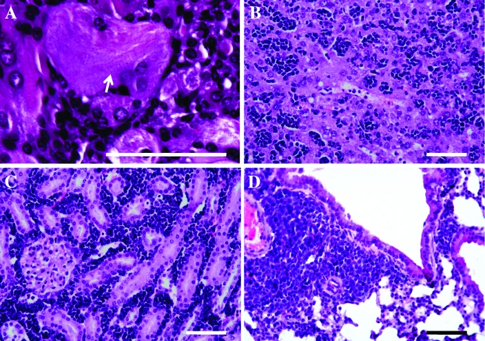
SCID mice provide an excellent platform for cancer research. Because of their lack of immunity, SCID mice readily succumb to infectious pathogens and therefore must be maintained in an SPF, barrier-protected environment. Although SPF and barrier facilities prevent infection, SCID mice remain prone to premature death due in part to a high prevalence of spontaneous thymic lymphomas. However, little is known about spontaneous nonthymic tumors in SCID mice. We therefore analyzed the incidence of nonthymic tumor in our defined-flora C.B-17/Icr-SCID/Sed mice and examined their histopathologic characteristics. We necropsied 1060 retired SCID breeders (506 males, 554 females; average ages of 325 and 320 d, respectively) and found that 24 mice had developed nonthymic tumors, yielding an incidence of 2.26% (1.78% in males; 2.71% in females). The incidence of nonthymic tumors was substantially lower than that of thymic lymphomas in our retired SCID breeders (12.3% in males; 4.15% in females). Based on histopathology, 9 nonthymic tumors in male SCID mice consisted of 4 salivary gland myoepiteliomas, 2 rhabdomyosarcomas, and 3 cases of leukemia involving multiple organs. Female SCID mice had 15 nonthymic tumors consisting of 8 mammary adenocarcinomas, 4 salivary gland myoepitheliomas, and 1 case each of leukemia, rhabdomyosarcoma, and fibrosarcoma. In addition, we tested in vivo transplantability and characterized the growth behavior of several of these tumors. To our knowledge, this report is the first comprehensive description of spontaneous nonthymic tumors, including 8 myoepitheliomas and 3 rhabdomyosarcomas, from the same SCID mouse colony.
Copyright 2011 by the American Association for Laboratory Animal Science
Figures
Similar articles
-
Existence of a threshold-like dose for gamma-ray induction of thymic lymphomas and no susceptibility to radiation-induced solid tumors in SCID mice.Mutat Res. 2007 Jun 1;619(1-2):124-33. doi: 10.1016/j.mrfmmm.2007.02.028. Epub 2007 Mar 4. Mutat Res. 2007. PMID: 17397880
-
Development of nonthymic lymphomas in thymectomized NFS mice exposed to split-dose X-irradiation.J Radiat Res. 1990 Dec;31(4):389-95. doi: 10.1269/jrr.31.389. J Radiat Res. 1990. PMID: 2098558
-
Spontaneous thymic lymphoma in severe combined immunodeficiency (SCID) mice engrafted with human peripheral blood lymphocytes.Vet Pathol. 1994 May;31(3):393-5. doi: 10.1177/030098589403100318. Vet Pathol. 1994. PMID: 8053140 No abstract available.
-
The relationship of pemphigus to neoplasia.J Am Acad Dermatol. 1990 Sep;23(3 Pt 1):498-502. doi: 10.1016/0190-9622(90)70249-h. J Am Acad Dermatol. 1990. PMID: 2212152 Review.
-
[Spontaneous tumors in rats of different lines].Vopr Onkol. 1976;22(8):98-110. Vopr Onkol. 1976. PMID: 793173 Review. Russian. No abstract available.
Cited by
-
Investigating circulating tumor cells and distant metastases in patient-derived orthotopic xenograft models of triple-negative breast cancer.Breast Cancer Res. 2019 Aug 28;21(1):98. doi: 10.1186/s13058-019-1182-4. Breast Cancer Res. 2019. PMID: 31462307 Free PMC article.
-
Barriers to generating PDX models of HPV-related head and neck cancer.Laryngoscope. 2017 Dec;127(12):2777-2783. doi: 10.1002/lary.26679. Epub 2017 May 31. Laryngoscope. 2017. PMID: 28561270 Free PMC article.
-
Ectopic TLX1 expression accelerates malignancies in mice deficient in DNA-PK.PLoS One. 2014 Feb 26;9(2):e89649. doi: 10.1371/journal.pone.0089649. eCollection 2014. PLoS One. 2014. PMID: 24586935 Free PMC article.
-
Newly Characterized Murine Undifferentiated Sarcoma Models Sensitive to Virotherapy with Oncolytic HSV-1 M002.Mol Ther Oncolytics. 2017 Sep 13;7:27-36. doi: 10.1016/j.omto.2017.09.003. eCollection 2017 Dec 15. Mol Ther Oncolytics. 2017. PMID: 29034313 Free PMC article.
-
Isolation and Characterization of a Novel Mammary Adenocarcinoma, MCa-P1362, with Hormone Receptor Expression, Human Epidermal Growth Factor Receptor 2 Positivity, and Enrichment in Cancer and Mesenchymal Stem Cells.Am J Pathol. 2024 Jun;194(6):1137-1153. doi: 10.1016/j.ajpath.2024.02.013. Am J Pathol. 2024. PMID: 38749609 Free PMC article.
References
-
- Booth CJ, Sundberg JP. 1996. Spontaneous neoplasms in a larger breeding colony of BALA/cJ and BALB/cByJ mice, p 51–65 Mohr U, Dungworth DL, Capen CC, Carlton WW, Sundberg JP, Ward JM. Pathobiology of the aging mouse. Washington (DC): ILSI Press
-
- Bosma GC, Custer RP, Bosma MJ. 1983. A severe combined immunodeficiency mutation in the mouse. Nature 301:527–530 - PubMed
Publication types
MeSH terms
Grants and funding
LinkOut - more resources
Full Text Sources