

Alterations in peripheral blood B-cell populations in SHIV89.6P-infected macaques (Macacca fascicularis)
- PMID: 21819698
- PMCID: PMC3123761
Alterations in peripheral blood B-cell populations in SHIV89.6P-infected macaques (Macacca fascicularis)
Abstract
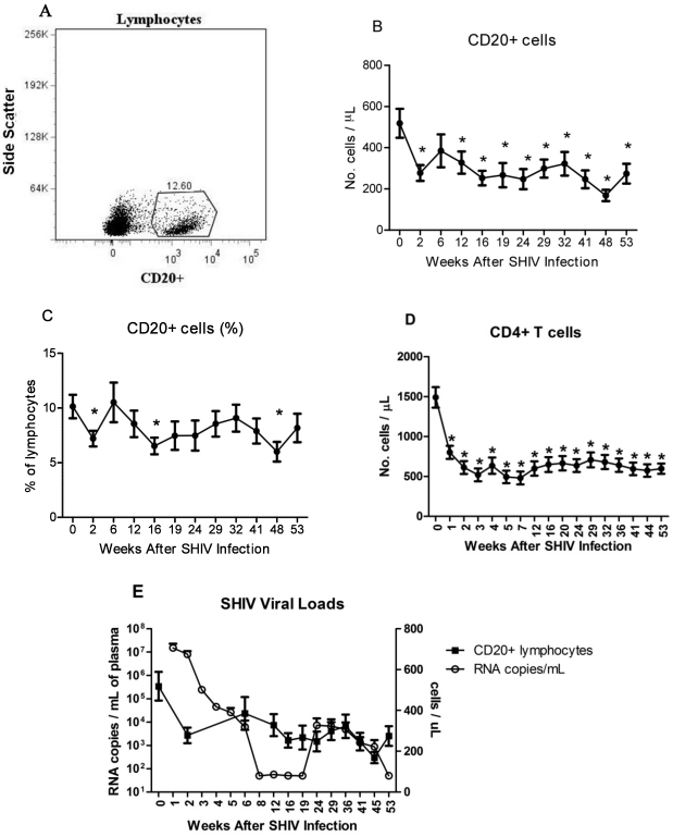
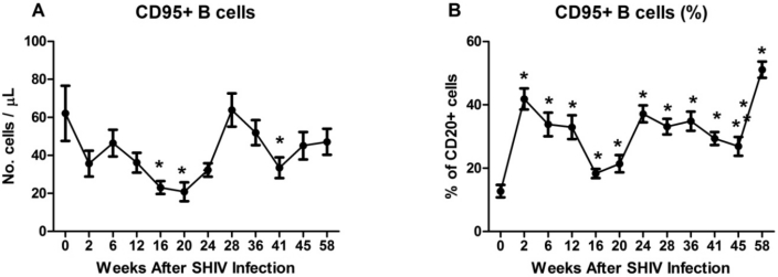
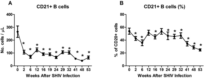
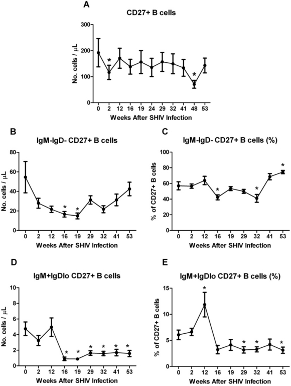
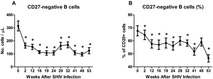
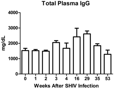
In addition to CD4+ T cell depletion, the B cell compartment of HIV-infected patients exhibits abnormalities, including deficits and diminished responses to ex vivo antigenic stimulation and in vivo vaccination. We used chimeric simian-human immunodeficiency virus (SHIV) infection of cynomolgus macaques to determine the dynamics of peripheral blood B cell alterations in this model of HIV infection. During the course of infection, we observed progressive loss of total and memory (CD27+) B cells, increased percentages of activated (CD95+) B cells, hypergammaglobulinemia, and deficits in the CD21+ B cell population. In addition, we noted declines in subsets of memory B cells, including both IgM+ and class-switched (IgD-IgM- CD27+) cells, with sustained deficits in the IgM+ memory (IgM+CD27+) B cell population. The similarity of the B cell alterations in these studies to those observed in HIV+ subjects supports the utility of the SHIV macaque model for examination of HIV-related B cell dysfunction.
Copyright 2011 by the American Association for Laboratory Animal Science
Figures
Similar articles
-
Vaccination with the Conserved Caveolin-1 Binding Motif in Human Immunodeficiency Virus Type 1 Glycoprotein gp41 Delays the Onset of Viral Infection and Provides Partial Protection in Simian/Human Immunodeficiency Virus-Challenged Cynomolgus Macaques.J Virol. 2018 Aug 29;92(18):e00370-18. doi: 10.1128/JVI.00370-18. Print 2018 Sep 15. J Virol. 2018. PMID: 29976675 Free PMC article.
-
Simian immunodeficiency virus SIVmac239 infection and simian human immunodeficiency virus SHIV89.6P infection result in progression to AIDS in cynomolgus macaques of Asian origin.J Gen Virol. 2016 Dec;97(12):3413-3426. doi: 10.1099/jgv.0.000641. Epub 2016 Oct 21. J Gen Virol. 2016. PMID: 27902330
-
Role of apoptosis induction in both peripheral lymph nodes and thymus in progressive loss of CD4+ cells in SHIV-infected macaques.AIDS Res Hum Retroviruses. 2000 Jan 1;16(1):9-18. doi: 10.1089/088922200309557. AIDS Res Hum Retroviruses. 2000. PMID: 10628812
-
Double-positive CD21+CD27+ B cells are highly proliferating memory cells and their distribution differs in mucosal and peripheral tissues.PLoS One. 2011 Jan 27;6(1):e16524. doi: 10.1371/journal.pone.0016524. PLoS One. 2011. PMID: 21304587 Free PMC article.
-
V3 loop-determined coreceptor preference dictates the dynamics of CD4+-T-cell loss in simian-human immunodeficiency virus-infected macaques.J Virol. 2005 Oct;79(19):12296-303. doi: 10.1128/JVI.79.19.12296-12303.2005. J Virol. 2005. PMID: 16160156 Free PMC article.
Cited by
-
Flow cytometric characterizations of leukocyte subpopulations in the peripheral blood of northern pig-tailed macaques (Macaca leonina).Dongwuxue Yanjiu. 2014 Nov 18;35(6):465-73. doi: 10.13918/j.issn.2095-8137.2014.6.465. Dongwuxue Yanjiu. 2014. PMID: 25465082 Free PMC article.
-
Pneumocystis infection and the pathogenesis of chronic obstructive pulmonary disease.Immunol Res. 2011 Aug;50(2-3):175-80. doi: 10.1007/s12026-011-8218-x. Immunol Res. 2011. PMID: 21717077 Free PMC article.
-
Vaccine-Induced Immunogenicity and Protection Against Pneumocystis Pneumonia in a Nonhuman Primate Model of HIV and Pneumocystis Coinfection.J Infect Dis. 2016 May 15;213(10):1586-95. doi: 10.1093/infdis/jiw032. Epub 2016 Jan 27. J Infect Dis. 2016. PMID: 26823337 Free PMC article.
-
Dynamics of memory B-cell populations in blood, lymph nodes, and bone marrow during antiretroviral therapy and envelope boosting in simian immunodeficiency virus SIVmac251-infected rhesus macaques.J Virol. 2012 Dec;86(23):12591-604. doi: 10.1128/JVI.00298-12. Epub 2012 Sep 12. J Virol. 2012. PMID: 22973034 Free PMC article.
References
-
- Amadori A, Zamarchi R, Ciminale V, Del Mistro A, Siervo S, Alberti A, Colombatti M, Chieco-Bianchi L. 1989. HIV1-specific B cell activation. A major constituent of spontaneous B cell activation during HIV1 infection. J Immunol 143:2146–2152 - PubMed
-
- Animal Welfare Act as Amended. 2007. 7 USC §2131-2159.
-
- Cagigi A, Nilsson A, De Milito A, Chiodi F. 2008. B cell immunopathology during HIV1 infection: lessons to learn for HIV1 vaccine design. Vaccine 26:3016–3025 - PubMed
-
- Chalifoux LV, King NW, Letvin NL. 1984. Morphologic changes in lymph nodes of macaques with an immunodeficiency syndrome. Lab Invest 51:22–26 - PubMed
Publication types
MeSH terms
Substances
Grants and funding
LinkOut - more resources
Full Text Sources
Research Materials