. 2011 Oct;179(4):2108-19.
doi: 10.1016/j.ajpath.2011.06.017. Epub 2011 Aug 3.

Phosphatase and tensin homolog on chromosome 10 is phosphorylated in primary effusion lymphoma and Kaposi's sarcoma

Affiliations

Phosphatase and tensin homolog on chromosome 10 is phosphorylated in primary effusion lymphoma and Kaposi's sarcoma

Debasmita Roy et al. Am J Pathol. 2011 Oct.

Abstract

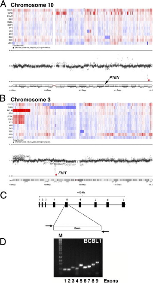
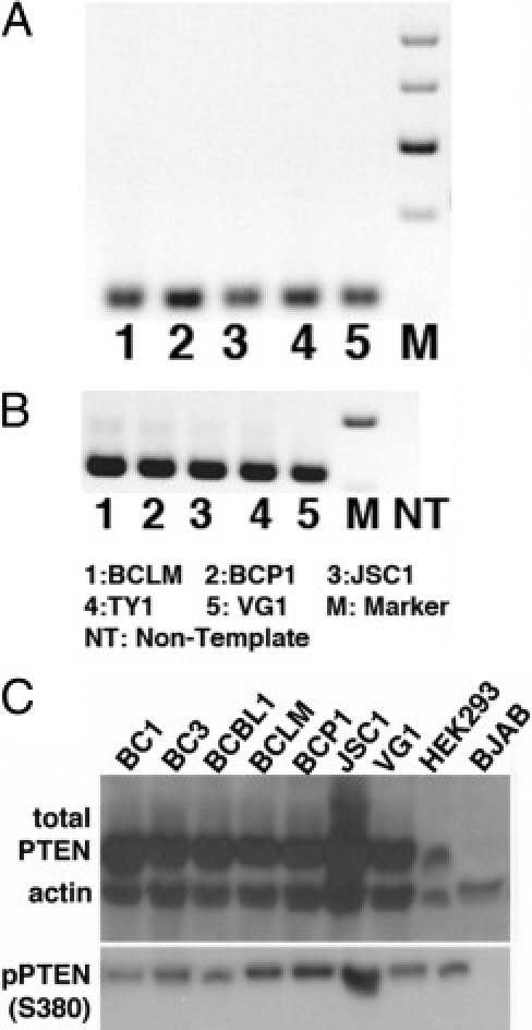
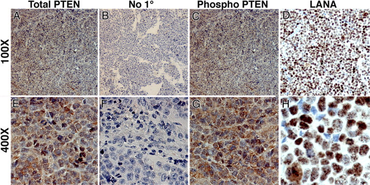
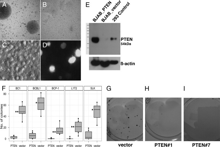
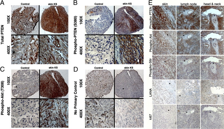
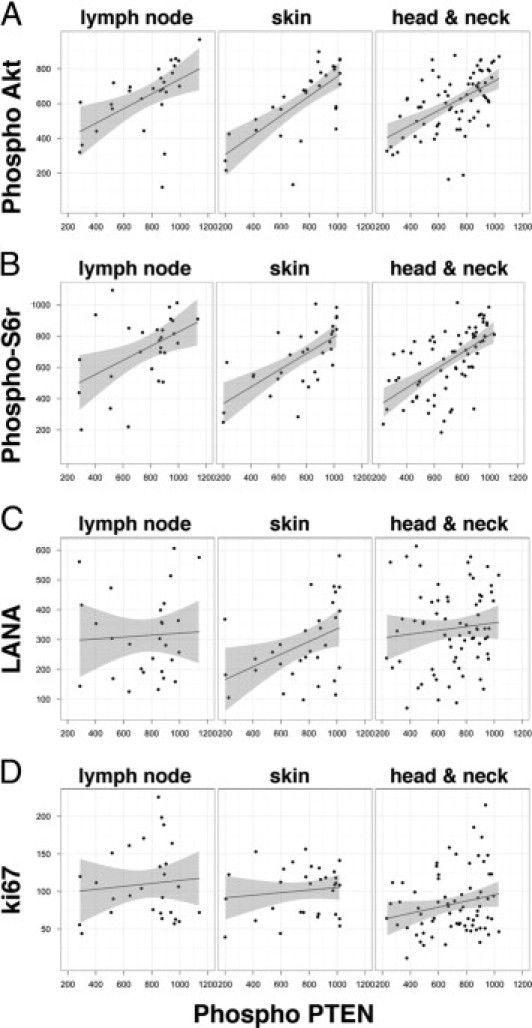
Primary effusion lymphoma (PEL) is a non-Hodgkin's B-cell lymphoma driven by Kaposi's sarcoma-associated herpesvirus. It is uniquely sensitive to mTOR, PI3K, and Akt inhibitors; however, the basis of this requirement for the mTOR pathway remains to be elucidated. The phosphatase and tensin homolog gene (PTEN) on chromosome 10 controls the first step in the phosphatidylinositol 3 kinase (PI3K)-Akt-mammalian target of rapamycin (mTOR) pathway and is genetically inactivated in many solid tumors. We find an absence of PTEN deletions, mutations, or protein mislocalization in PEL. However, we find consistent hyperphosphorylation at serine 380 of PTEN, which is an inactivating modification, in PEL cell lines and in tumor xenografts. We also evaluated a large tissue microarray of Kaposi's sarcoma biopsies and observed concordant high levels of phospho-PTEN, phospho-Akt, and phospho-S6 ribosomal protein. Reintroduction of PTEN into PEL inhibited colony formation in soft agar, verifying the functional dependence of PEL on PI3K signaling. This was also true for PEL cell lines that carried mutant p53 and for KS-like cell lines. Activating PTEN in these cancers may yield a new treatment strategy for PEL, KS, and similar PTEN wild-type lymphomas.


Figures

Figure 1
Figure 1
PTEN is wild type in PEL. A and B: Using an Affymetrix 500K single nucleotide polymorphism array, we observed that the majority of PELs do not show genomic gain or loss at the PTEN locus on chromosome 10 (A). Chromosome 3 served as control; distinct loss was noted at the FHIT locus, a known common fragile site gene (B). Heat maps indicate loss (blue) and gain (red). The dot profiles show the distribution of markers along the chromosome; an asterisk marks a small region that is specifically lost in an otherwise normal chromosomal region. On chromosome 10 (A), this does not correspond to any known transcripts; on chromosome 3 (B), it corresponds to FHIT. The labeled cytobands indicate location of PTEN (A) and FHIT (B). C and D: Genomic DNA was isolated from individual PEL cell lines and each of the exons was amplified using specific primers (listed in Table 2). C: Schematic of the organization of the primers used. D: A representative PCR gel showing detection of all nine exons in BCBL-1 cells.
Figure 2
Figure 2
PTEN is phosphorylated at the serine 380 site in PEL. RT-PCR was performed to determine PTEN mRNA expression in PEL; representative gels are shown for PTEN (A) and GAPDH (B). NT, nontemplate control. C: Immunoblots were performed on protein isolated from whole-cell lysate; representative immunoblots are shown for total PTEN and phospho-PTEN (S380). β-actin was used as loading control. Equivalent amounts of protein were loaded in each case, except for HEK293, which served to indicate the molecular weight of PTEN.
Figure 3
Figure 3
Immunohistochemical analysis of PEL xenograft tumors shows similar localization of total PTEN and phospho-PTEN. BC-1 xenograft tumor sections were stained for total PTEN (A and E), phospho-PTEN (S380) (C and G), and LANA (D and H) (red; NovaRed substrate). Sections were counterstained using hematoxylin (blue), which identifies the nucleus. Sections shown were incubated with dilution buffer either with primary antibody at 1:100 dilution (A, C, D, E and H) or without primary antibody (B and F). No red staining was observed in the no-primary-antibody control (B and D). Original magnification: ×100 (AD); ×400 (EH).
Figure 4
Figure 4
Growth suppression of BCP-1 and SLK cells upon overexpression of PTEN. BCP1 cells colonies formed in soft agar after vector-only nucleofection (A), but no colonies formed on nucleofection of human PTEN clone 1 (pDD1540) (B). GFP positive control for nucleofection is shown in bright light (C) and fluorescence (D) fields. E: Expression of protein in PTEN-null BJAB cells on expression of the hPTEN only and not the vector control. F: Quantification of colonies observed in the PEL cell lines BC1, BCBL1, and BCP1 (P = 0.003, 0.02, and 0.005, respectively) and in the KS-like cell lines L1T2 and SLK (P = 0.003 and 0.03, respectively). *P ≤ 0.05; **P ≤ 0.005. G–I: Representative images of colonies upon overexpression of hPTEN, clones 1 (H) and 7 (I) compared with vector only (G) in SLK cells.
Figure 5
Figure 5
Expression of PI3K activation markers and phospho-PTEN (S380) in KS primary biopsies. Immunohistochemistry showed the presence of total PTEN (A), phospho-PTEN (S380) (B), phospho-Akt (T308), a PI3K-dependent site (C), and no-primary-antibody negative control (D). Original magnification: ×100 (upper row); ×400 (lower row). In each panel (A–D), images at the left are of control lung tissue, which stains positive only for PTEN but not for phospho-PTEN (S380) or phospho-Akt (T308); images at the right are of a representative skin biopsy, in which the same tissue section is positive for total PTEN, phospho-PTEN (S380), and phospho-Akt (T308). E: Staining of a second TMA incorporating AIDS-KS from tissues of three different origins for phospho-PTEN (S380), PI3K activation markers phospho-Akt (T308) and phospho-S6r (S235/236), KSHV marker LANA, and proliferation marker Ki67. Sections were selected such that a region of the biopsy was negative (asterisks), in an otherwise positive section, serving as an internal control. Original magnification, ×100.
Figure 6
Figure 6
Significant correlation of phospho-Akt (T308) and phospho-S6 ribosomal protein (S235/236) with phospho-PTEN (S380) in KS. The square root of the intensity count for phospho-Akt (A), phospho-S6r (B), LANA (C), and Ki67 (D), is plotted on the vertical axis and that for phospho-PTEN is plotted on the horizontal axis for KS sections from three body sites: lymph node, oral, and skin. The solid line indicates the linear regression; the shaded region indicates the 95% confidence interval of the fit. There was a significant correlation of both phospho-Akt (T308) and phospho-S6r (S235/236) with phospho-PTEN (S380), with P values of 3.09 × 10⁻¹³ and 2.6 × 10⁻¹³ respectively. There was no significant correlation between LANA positivity (denoting viral infection) and Ki67 (denoting active proliferation) (P > 0.05).

Similar articles

Cited by

References

    1. Cesarman E., Chang Y., Moore P.S., Said J.W., Knowles D.M. Kaposi's sarcoma-associated herpesvirus-like DNA sequences in AIDS-related body-cavity-based lymphomas. N Engl J Med. 1995;332:1186–1191. - PubMed
    1. Matsushima A.Y., Strauchen J.A., Lee G., Scigliano E., Hale E.E., Weisse M.T., Burstein D., Kamel O., Moore P.S., Chang Y. Posttransplantation plasmacytic proliferations related to Kaposi's sarcoma-associated herpesvirus. Am J Surg Pathol. 1999;23:1393–1400. - PubMed
    1. Dotti G., Fiocchi R., Motta T., Facchinetti B., Chiodini B., Borleri G.M., Gavazzeni G., Barbui T., Rambaldi A. Primary effusion lymphoma after heart transplantation: a new entity associated with human herpesvirus-8. Leukemia. 1999;13:664–670. - PubMed
    1. Nador R.G., Cesarman E., Chadburn A., Dawson D.B., Ansari M.Q., Sald J., Knowles D.M. Primary effusion lymphoma: a distinct clinicopathologic entity associated with the Kaposi's sarcoma-associated herpes virus. Blood. 1996;88:645–656. - PubMed
    1. Horenstein M.G., Nador R.G., Chadburn A., Hyjek E.M., Inghirami G., Knowles D.M., Cesarman E. Epstein-Barr virus latent gene expression in primary effusion lymphomas containing Kaposi's sarcoma-associated herpesvirus/human herpesvirus-8. Blood. 1997;90:1186–1191. - PubMed

Publication types

MeSH terms