. 2012 Feb 10;157(3):354-65.
doi: 10.1016/j.jconrel.2011.07.029. Epub 2011 Jul 24.

Robust IgG responses to nanograms of antigen using a biomimetic lipid-coated particle vaccine

Affiliations

Robust IgG responses to nanograms of antigen using a biomimetic lipid-coated particle vaccine

Anna Bershteyn et al. J Control Release. .

Abstract

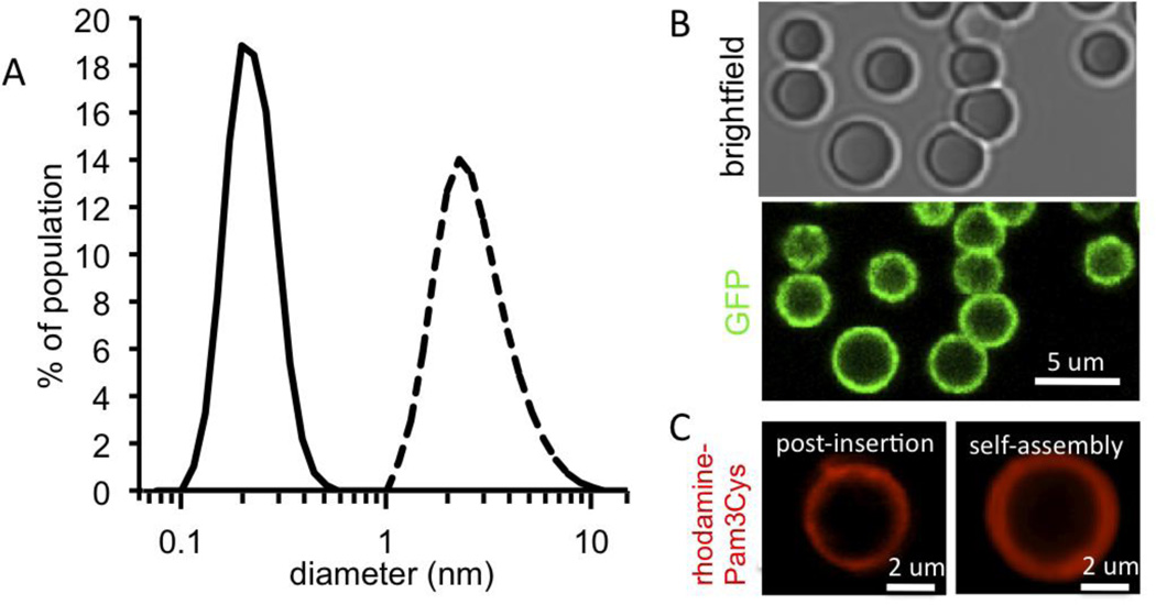
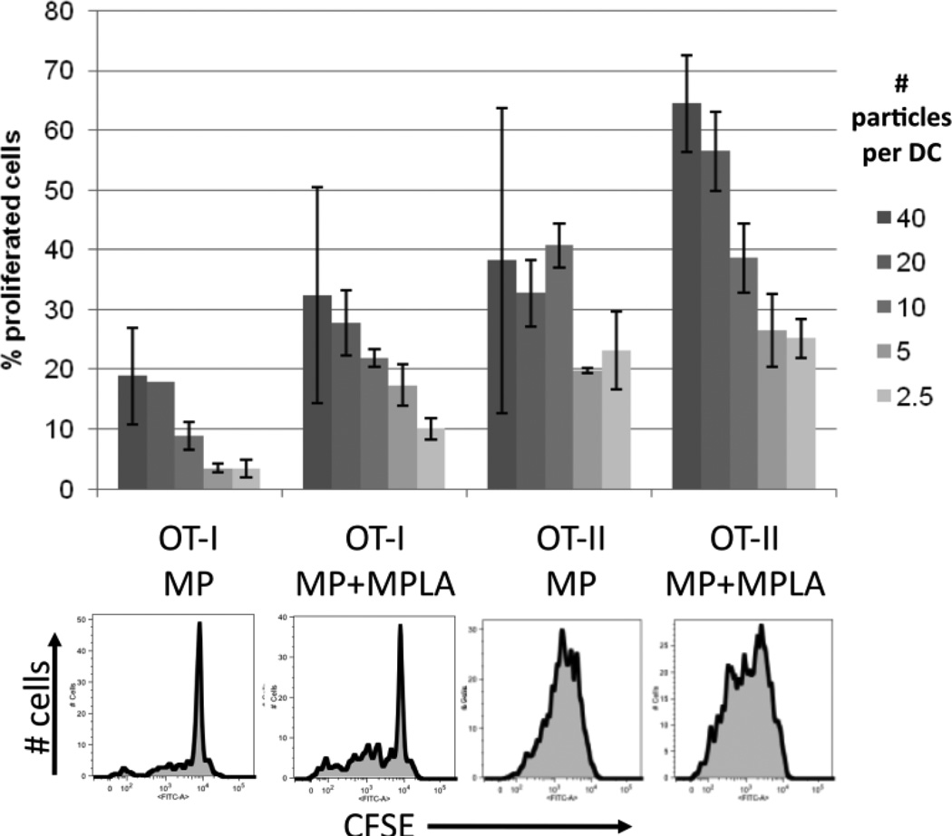
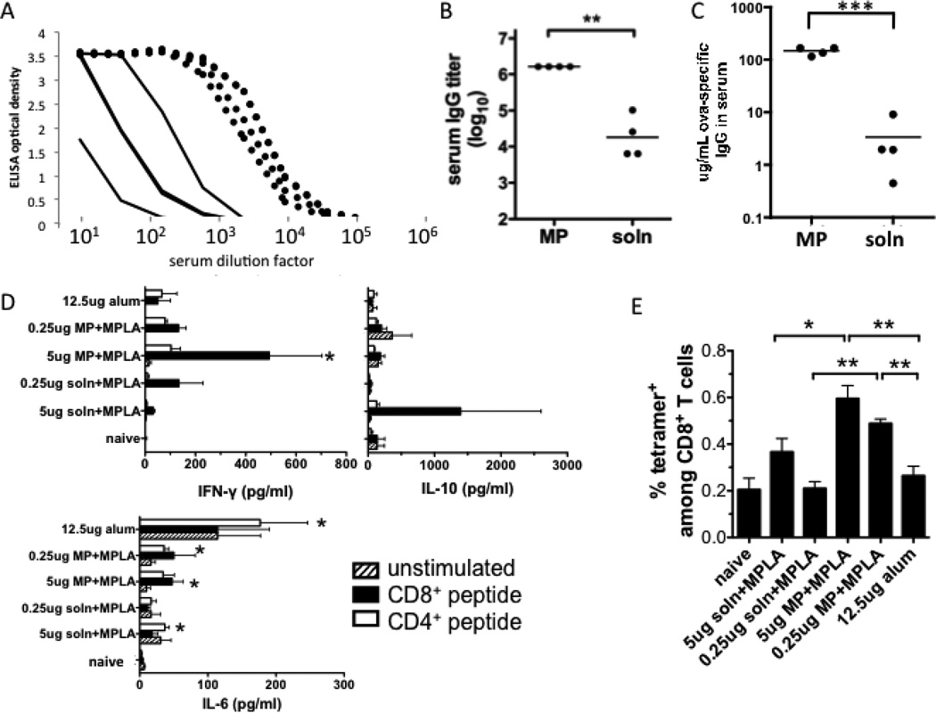
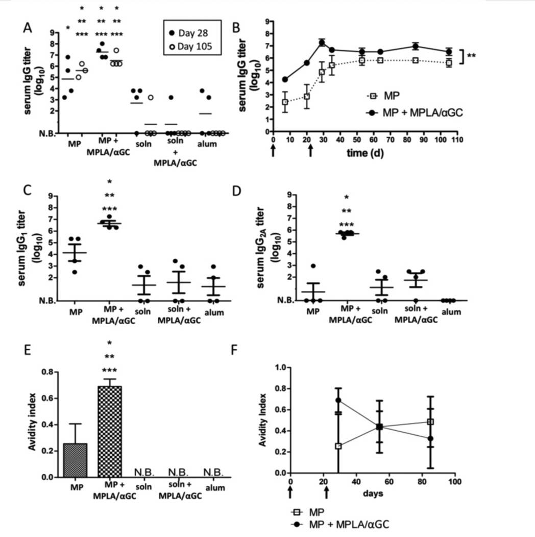
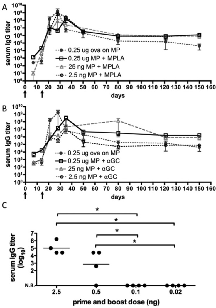
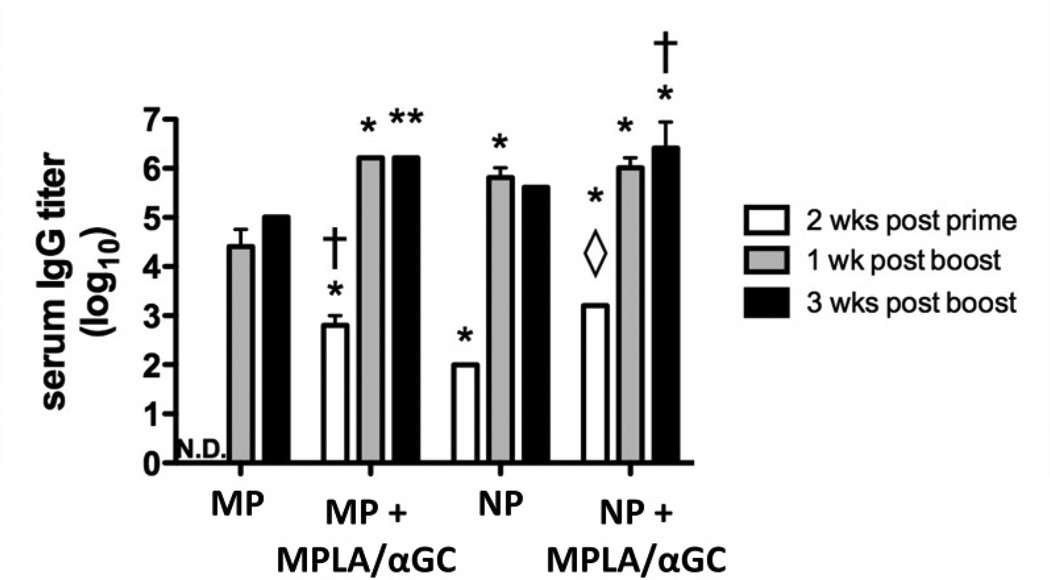
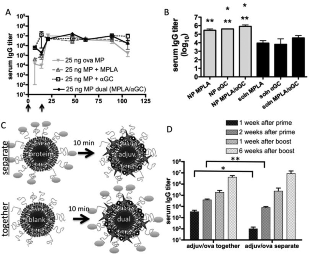
New subunit vaccine formulations with increased potency are of interest to improve immune responses against poorly immunogenic antigens, to avoid vaccine shortages in pandemic situations, and to promote dose-sparing of potent adjuvant molecules that can cause unacceptable side effects in prophylactic vaccination. Here we report strong class-switched, high avidity humoral immune responses elicited by a vaccine system based on poly(lactide-co-glycolide) micro- or nano-particles enveloped by PEGylated phospholipid bilayers, with protein antigens covalently anchored to the lipid surface and lipophilic adjuvants inserted in the bilayer coating. Strikingly, these particles elicited high endpoint antigen-specific IgG titers (>10(6)) sustained for over 100 days after two immunizations with as little as 2.5 ng of antigen. At such low doses, the conventional adjuvant alum or the molecular adjuvants monophosphoryl lipid A (MPLA) or α-galactosylceramide (αGC) failed to elicit responses. Co-delivery of antigen with MPLA or αGC incorporated into the particle bilayers in a pathogen-mimetic fashion further enhanced antibody titers by ~12-fold. MPLA provided the highest sustained IgG titers at these ultra-low antigen doses, while αGC promoted a rapid rise in serum IgG after one immunization, which may be valuable in emergencies such as disease pandemics. The dose of αGC required to boost the antibody response was also spared by particulate delivery. Lipid-enveloped biodegradable micro- and nano-particles thus provide a potent dose-sparing platform for vaccine delivery.


Figures

Figure 1
Figure 1. Synthesis of lipid-enveloped micro- or nano-particles with surface-displayed antigen and molecular adjuvants
(A) Light scattering analysis of purified particle size distributions for microparticles (dashed line) or nanoparticles (solid line) synthesized by homogenization or sonication, respectively, to disperse lipid/polymer emulsion during particle synthesis. (B) Confocal imaging of lipid-enveloped microparticles bearing ~7×10⁴ green fluorescent protein molecules per particle (green, GFP intrinsic fluorescence). (C) Confocal imaging of microparticles modified with rhodamine-labeled Pam3Cys (red fluorescence, lipid-like TLR-2 agonist) incorporated via post-insertion or through self-assembly during particle synthesis.
Figure 2
Figure 2. Priming of naïve CD4+ or CD8+ T-cells by antigen-conjugated lipid-enveloped particles
Primary splenic DCs were incubated with ova-conjugated microparticles (with or without post-inserted MPLA) for 3 hrs, then co-cultured with naïve CFSE-labeled OT-I (CD8+) or OT-II (CD4+) T-cells. Proliferation of T-cells was assessed after 3 days by flow cytometry. Shown are representative flow histograms (10:1 particle:DC ratio) and mean percentages of proliferated cells from triplicate wells (± St. dev.). The maximum particle:DC ratio (40:1) corresponds to a total dose of 2.6 ng ova in the 200 µL culture.
Figure 3
Figure 3. Serum IgG responses to particle-delivered or soluble ova at a modest but conventional dose of 0.5 µg ova
(A–C) BALB/c mice were immunized s.c. with 500 ng of ova in solution or displayed on lipid-coated microparticles and boosted on day 21 with the same formulations. Shown are analyses of sera collected on day 28: (A) Total anti-ova IgG ELISA on serum from mice immunized with ova-particles (dotted lines) or ova solution (solid lines); (B) Endpoint total IgG titers (**, P < 0.01) (C) total ova-specific IgG concentration in sera (***, P<0.0001). (D, E) C57Bl/6 mice were immunized on day 0 and day 21 s.c. with indicated doses of ova with alum, ova-particles with MPLA, or ova solution mixed with MPLA (2.5 µg MPLA in all cases). 7 days after boosting, splenocytes were collected and restimulated ex vivo with immunodominant CD8+ or CD4+ ova peptides for analysis of cytokine production (D) and frequencies of ova-specific CD8+ T-cells in spleens were analyzed by peptide-MHC tetramer staining and flow cytometry (E). (D, *, P < 0.05 relative to naïve mice; E, *, P < 0.05; **, P < 0.01).
Figure 4
Figure 4. Serum IgG responses elicited by lipid-coated particles vs. conventional adjuvants at limiting antigen doses
Groups of BALB/c mice (n = 4) were immunized s.c. with 10 ng ova displayed on microparticles (“MP”), dissolved in saline (“soln”), mixed with alum, or mixed with MPLA and αGC; animals were boosted on day 21 with the same formulations. In both the particle-displayed and soluble adjuvant cases, equimolar quantities of 1.3 µg MPLA and 600 ng αGC were used. (A) Post-boost peak (day 28) and late (day 105) endpoint titers from individual mice. (B) Mean endpoint titers (±SEM) for particle immunizations over time (**, P=0.0053). (C, D) Endpoint IgG1 (C) and IgG2A titers at day 28. (E, F) Avidity of ova-specific IgG in each group measured at day 28 for all groups (E) or for the particle-immunized groups over time (F). (N.B.: No binding detected. *, **, *** in panels A, C–E: P < 0.05 relative to soln+MPLA/αGC, soln, or alum at the same time point, respectively).
Figure 5
Figure 5. IgG responses following dose sparing immunizations with lipid-coated particle immunogens
Groups of C57Bl/6 mice (n = 3) were immunized with lipid-coated microparticles delivering the indicated dose of ova and boosted on day 14. The particle-only conditions (black circles) carried ova alone; otherwise, 13 µg MPLA or 6 µg αGC were added via the post-insertion method to the antigen-loaded particles. Shown are mean endpoint titers (±SEM) for dose titrations of particles carrying ova and (A) MPLA or (B) αGC. (C) Groups of BALB/c mice (n = 4) were immunized with diminishing doses of ova co-displayed with 1.3 µg MPLA and boosted on day 14 to determine the minimum dose capable of eliciting measurable antibody responses. Post-boost peak (day 28) endpoint titers are shown for individual mice (*, P < 0.05).
Figure 6
Figure 6. Comparison of adjuvant effect of lipid-enveloped microparticles vs. nanoparticles
Groups of BALB/c mice (n = 3) were immunized s.c. with 10 ng ova displayed on microparticles (“MP”) or nanoparticles (“NP”), and boosted on day 14. For comparison, particles from the same syntheses were loaded with 1.3 µg MPLA and 600 ng αGC via the post-insertion method. Bars show mean endpoint titers ±SEM. N.D., no antigen-specific IgG detected above background (*, P < 0.001 vs. MP; **, P < 0.01 vs. MP; ◊, P < 0.01 vs. NP; †, P < 0.05 vs. NP; all comparisons made by Bonferroni post-tests at the same time point).
Figure 7
Figure 7. Analysis of synergy between MPLA and αGC in particle vaccine responses
Groups of BALB/c mice (n = 4) were immunized s.c. with ova displayed on microparticles and boosted on day 21; endpoint total IgG titers were determined and shown are means ± SEM. (A) Mice were immunized with 25 ng ova and 1.3 µg MPLA and/or 600 ng αGC co-loaded onto microparticles. (B) Mice were immunized with 10 ng ova-conjugated nanoparticles co-loaded with MPLA, αGC, or both adjuvants, and compared to mice given the same doses of the adjuvant molecules injected in soluble form 10 min before injection of the antigen-loaded particles at the same site. Titers were assessed on day 28 (*, P < 0.05 vs. soln MPLA; **, P < 0.05 vs. soln αGC). (C, D) Mice were immunized with microparticles displaying 10 ng ova, followed 10 minutes later by microparticles displaying 1.3 µg MPLA and 600 ng αGC injected at the same site (C, “separate”). For comparison, mice received an equivalent number of blank microparticles, followed 10 minutes later by microparticles co-displaying 10 ng ova, 1.3 µg MPLA, and 600 ng αGC (C, “together”). *, P = 0.0284; **, P = 0.0070.
Figure 8
Figure 8. Dose sparing of molecular adjuvants by lipid-coated particles
Groups of BALB/c mice (n = 4) were immunized s.c. with 2 ng ova displayed on microparticles co-loaded with the indicated quantities of αGC via the post-insertion method (αGC on particles). A second group of mice was immunized by injecting the indicated doses of αGC followed 10 min later by 2 ng ova-microparticles at the same site (αGC solution). Shown are endpoint total IgG titers for individual mice two weeks after a single immunization (*, P < 0.05).

References

    1. Bachmann MF, Rohrer UH, Kundig TM, Burki K, Hengartner H, Zinkernagel RM. The Influence of Antigen Organization on B-Cell Responsiveness. Science. 1993;262:1448–1451. - PubMed
    1. Chackerian B, Lowy DR, Schiller JT. Conjugation of a self-antigen to papillomavirus-like particles allows for efficient induction of protective autoantibodies. Journal of Clinical Investigation. 2001;108:415–423. - PMC - PubMed
    1. Dintzis HM, Dintzis RZ, Vogelstein B. Molecular Determinants of Immunogenicity - Immunon Model of Immune-Response. Proceedings of the National Academy of Sciences of the United States of America. 1976;73:3671–3675. - PMC - PubMed
    1. Gamvrellis A, Leong D, Hanley JC, Xiang SD, Mottram P, Plebanski M. Vaccines that facilitate antigen entry into dendritic cells. Immunology and Cell Biology. 2004;82:506–516. - PubMed
    1. Kovacsovicsbankowski M, Clark K, Benacerraf B, Rock KL. Efficient Major Histocompatibility Complex Class-I Presentation of Exogenous Antigen Upon Phagocytosis by Macrophages. Proceedings of the National Academy of Sciences of the United States of America. 1993;90:4942–4946. - PMC - PubMed

Publication types

MeSH terms