. 2011 Oct 15;51(8):1492-500.
doi: 10.1016/j.freeradbiomed.2011.07.004. Epub 2011 Jul 19.

Role of VPO1, a newly identified heme-containing peroxidase, in ox-LDL induced endothelial cell apoptosis

Affiliations

Role of VPO1, a newly identified heme-containing peroxidase, in ox-LDL induced endothelial cell apoptosis

Yong-Ping Bai et al. Free Radic Biol Med. .

Abstract

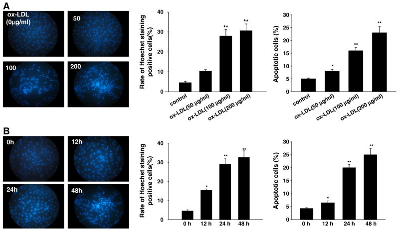
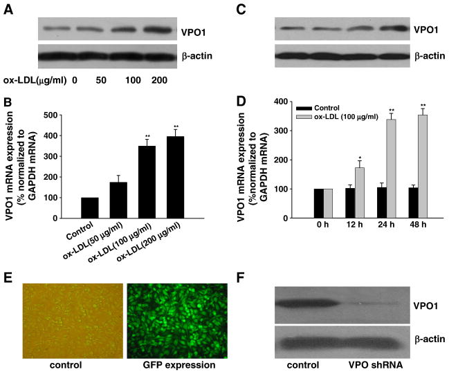
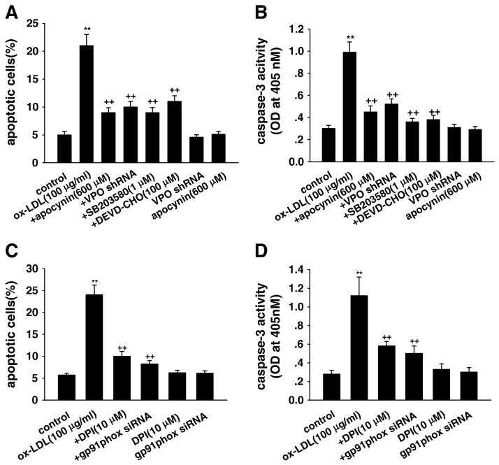
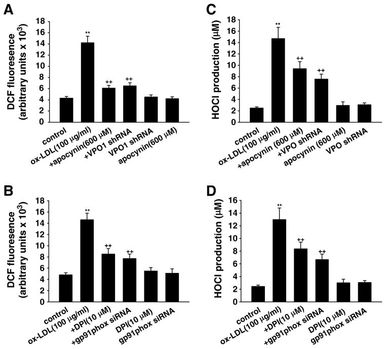
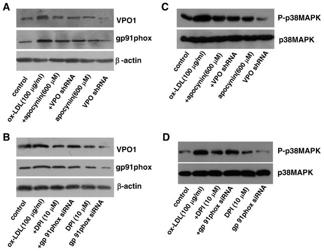
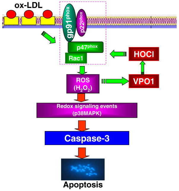
Myeloperoxidase (MPO) is an important enzyme involved in the genesis and development of atherosclerosis. Vascular peroxidase 1 (VPO1) is a newly discovered member of the peroxidase family that is mainly expressed in vascular endothelial cells and smooth muscle cells and has structural characteristics and biological activity similar to those of MPO. Our specific aims were to explore the effects of VPO1 on endothelial cell apoptosis induced by oxidized low-density lipoprotein (ox-LDL) and the underlying mechanisms. The results showed that ox-LDL induced endothelial cell apoptosis and the expression of VPO1 in endothelial cells in a concentration- and time-dependent manner concomitant with increased intracellular reactive oxygen species (ROS) and hypochlorous acid (HOCl) generation, and up-regulated protein expression of the NADPH oxidase gp91(phox) subunit and phosphorylation of p38 MAPK. All these effects of ox-LDL were inhibited by VPO1 gene silencing and NADPH oxidase gp91(phox) subunit gene silencing or by pretreatment with the NADPH oxidase inhibitor apocynin or diphenyliodonium. The p38 MAPK inhibitor SB203580 or the caspase-3 inhibitor DEVD-CHO significantly inhibited ox-LDL-induced endothelial cell apoptosis, but had no effect on intracellular ROS and HOCl generation or the expression of NADPH oxidase gp91(phox) subunit or VPO1. Collectively, these findings suggest for the first time that VPO1 plays a critical role in ox-LDL-induced endothelial cell apoptosis and that there is a positive feedback loop between VPO1/HOCl and the now-accepted dogma that the NADPH oxidase/ROS/p38 MAPK/caspase-3 pathway is involved in ox-LDL-induced endothelial cell apoptosis.


Figures

Fig. 1
Fig. 1
(A) Concentration response and (B) time course of ox-LDL-induced apoptosis in cultured HUVECs. (A) The endothelial cells were exposed to ox-LDL at various concentrations (50, 100, and 200 μg/ml) for 24 h. (B) The cells were exposed to ox-LDL (100 μg/ml) for 0, 12, 24, and 48 h. Apoptosis was determined by Hoechst 33342 staining and annexin V–PI double-staining assay (flow cytometry). Data are expressed as means±SEM, n=6 each, performed in triplicate. Compared with control (0 μg/ml ox-LDL) or 0 h, *P<0.05; **P<0.01.
Fig. 2
Fig. 2
Expression of VPO1 in response to ox-LDL in HUVECs and effect of knockdown of VPO1 by shRNA. (A, B, C and D) Ox-LDL up-regulated the expression of VPO1 (both mRNA and protein, analyzed by real-time PCR and Western blot, respectively) in a time- and concentration-dependent manner. (E) GFP immunofluorescence staining of the cells transfected with VPO1-shRNA. (F) VPO1-shRNA successfully decreased VPO1 protein expression. Data are expressed as means±SEM, n=6 each, performed in triplicate. Compared with control (0μg/ml ox-LDL) or 0 h, *P<0.05; **P<0.01.
Fig. 3
Fig. 3
Effects of VPO1 knockdown on apoptosis of HUVECs induced by ox-LDL. (A and C) Cell apoptosis analysis by flow cytometry. (B and D) Caspase-3 activity. Control, wild-type cells were treated with 0 μg/ml ox-LDL for 24 h; ox-LDL, wild-type cells were treated with 100 μg/ml ox-LDL for 24 h; +DPI, cells were pretreated with 10 μM DPI (the specific NADPH oxidase inhibitor) for 1 h before ox-LDL exposure; +apocynin, cells were pretreated with 600 μM apocynin for 1 h before ox-LDL exposure; +gp91phox siRNA, after successful gp91phox siRNA transfection, cells were cultured in DMEM containing 100 μg/ml ox-LDL for 24 h; +VPO shRNA, after successful VPO1 shRNA transfection, cells were cultured in DMEM containing 100 μg/ml ox-LDL for 24 h; +SB203580, cells were pretreated with 1 μM SB203580 (the specific p38 MAPK inhibitor) for 1 h before ox-LDL exposure; +DEVD-CHO, cells were pretreated with 100 μM DEVD-CHO (the specific caspase-3 inhibitor) for 1 h before ox-LDL exposure; gp91phox siRNA, cells with successful gp91phox siRNA transfection were incubated in DMEM containing 1% calf serum for 24 h; VPO shRNA, cells with successful VPO1 shRNA transfection were incubated in DMEM containing 1% calf serum for 24 h; DPI, cells were cultured in DMEM containing 10 μM DPI for 24 h; apocynin, cells were cultured in DMEM containing 600 μM apocynin for 24 h. **P<0.01 vs control (0 μg/ml ox-LDL), ++P<0.01 vs ox-LDL (100 μg/ml). Data are expressed as means±SEM, n=6 each, performed in triplicate.
Fig. 4
Fig. 4
The role of intracellular ROS and HOCl in ox-LDL-induced apoptosis in HUVECs. (A and B) Intracellular ROS level was determined by fluorescent DCF. (C and D) HOCl production was determined by TMB assay. Control, wild-type cells were incubated in 10 mM phosphate buffer containing 5 mM taurine for 30 min; ox-LDL (100 μg/ml), wild-type cells were cultured in 10 mM phosphate buffer containing 5 mM taurine and ox-LDL (100 μg/ml) for 30 min; +DPI, cells were pretreated with 10 μM DPI ( the specific NADPH oxidase inhibitor) for 1 h before ox-LDL exposure; +apocynin, cells were pretreated with 600 μM apocynin for 1 h before ox-LDL exposure; +gp91phox siRNA, after successful gp91phox siRNA transfection, cells were cultured in 10 mM phosphate buffer containing 5 mM taurine and ox-LDL (100 μg/ml) for 30 min; +VPO shRNA, after successful VPO1 shRNA transfection, cells were cultured in 10 mM phosphate buffer containing 5 mM taurine and ox-LDL (100 μg/ml) for 30 min; gp91phox siRNA, cells with successful gp91phox siRNA transfection were incubated in 10 mM phosphate buffer containing 5 mM taurine for 30 min; VPO shRNA, cells with successful VPO shRNA transfection were incubated in 10 mM phosphate buffer containing 5 mM taurine for 30 min; DPI, cells were cultured in 10 mM phosphate buffer containing 5 mM taurine and 10 μM DPI for 30 min; apocynin, cells were cultured in 10 mM phosphate buffer containing 5 mM taurine and 600 μM apocynin for 30 min. **P<0.01 vs control (0 μg/ml ox-LDL), ++P<0.01 vs ox-LDL (100 μg/ml). Data are expressed as means±SEM, n=6 each, performed in triplicate.
Fig. 5
Fig. 5
Relationship between VPO1 and the NADPH oxidase/p38 MAPK pathway in ox-LDL-induced apoptosis in HUVECs. (A and B) The protein levels of VPO1 and NADPH oxidase subunit gp91phox. (C and D) The protein levels of (phosphorylated) p38 MAPK. Control, endothelial cells were incubated in DMEM containing 1% calf serum for 24 h; ox-LDL (100 μg/ml), endothelial cells were cultured in DMEM containing 100 μg/ml ox-LDL for 24 h; +DPI, cells were pretreated with 10 μM DPI (the specific NADPH oxidase inhibitor) for 1 h before ox-LDL exposure; +apocynin, cells were pretreated with 600 μM apocynin for 1 h before ox-LDL exposure; +gp91phox siRNA, after successful gp91phox siRNA transfection, cells were cultured in DMEM containing 100 μg/ml ox-LDL for 24 h; +VPO shRNA, after successful VPO1 shRNA transfection, cells were cultured in DMEM containing 100 μg/ml ox-LDL for 24 h; gp91phox siRNA, cells with successful gp91phox siRNA transfection were incubated in DMEM containing 1% calf serum for 24 h; VPO shRNA, cells with successful VPO1 shRNA transfection were incubated in DMEM containing 1% calf serum for 24 h; DPI, cells were cultured in DMEM containing 10 μM DPI for 24 h; apocynin, cells were cultured in DMEM containing 600 μM apocynin for 24 h. n=6 each, performed in triplicate.
Fig. 6
Fig. 6
The proposed pathway of VPO1 mediation of ox-LDL-induced endothelial cell apoptosis. Ox-LDL up-regulates the expression of NADPH oxidase subunit gp91phox and consequently increases intracellular ROS production and VPO1 expression as well as HOCl production. There is a positive feedback loop between VPO1/HOCl and the NADPH oxidase/ROS pathway, which in turn activates the p38 MAPK/caspase-3-dependent signaling pathway to mediate ox-LDL-induced endothelial cell apoptosis.

References

    1. Malle E, Waeg G, Schreiber R, Gröne EF, Sattler W, Gröne HJ. Immunohistochemical evidence for the myeloperoxidase/H2O2/halide system in human atherosclerotic lesions. Eur J Biochem. 2000;267:4495–4503. - PubMed
    1. Podrez EA, Abu-Soud HM, Hazen SL. Myeloperoxidase-generated oxidants and atherosclerosis. Free Radic Biol Med. 2000;28:1717–1725. - PubMed
    1. Stocker R, Huang A, Jeranian E, Hou JY, Wu TT, Thomas SR, Keaney JF., Jr Hypochlorous acid impairs endothelium-derived nitric oxide bioactivity through a superoxide-dependent mechanism. Arterioscler Thromb Vasc Biol. 2004;24:2028–2033. - PubMed
    1. Woods AA, Linton SM, Davies MJ. Detection of HOCl-mediated protein oxidation products in the extracellular matrix of human atherosclerotic plaques. Biochem J. 2003;370:729–735. - PMC - PubMed
    1. Malle E, Marsche G, Arnhold J, Davies MJ. Modification of low-density lipoprotein by myeloperoxidase-derived oxidants and reagent hypochlorous acid. Biochim Biophys Acta. 2006;1761:392–415. - PubMed

Publication types

MeSH terms