. 2011 Aug 12;89(2):219-30.
doi: 10.1016/j.ajhg.2011.06.013. Epub 2011 Aug 4.

KIF1A, an axonal transporter of synaptic vesicles, is mutated in hereditary sensory and autonomic neuropathy type 2

Affiliations

KIF1A, an axonal transporter of synaptic vesicles, is mutated in hereditary sensory and autonomic neuropathy type 2

Jean-Baptiste Rivière et al. Am J Hum Genet. .

Abstract

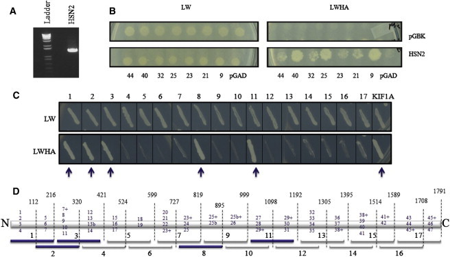
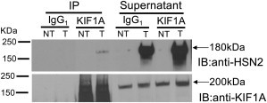
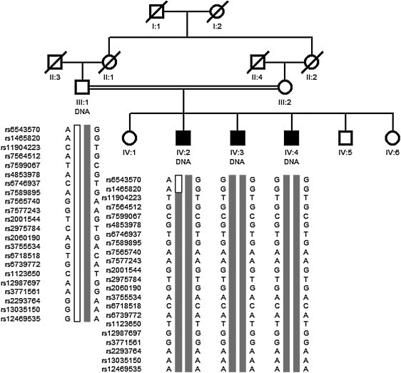
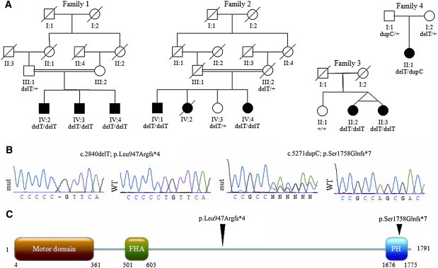
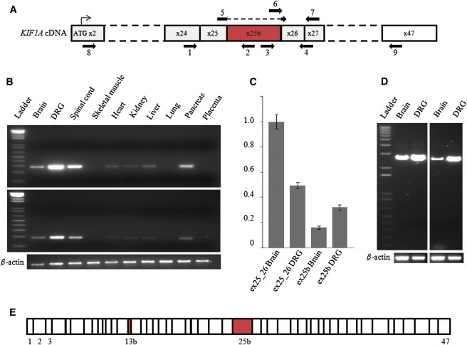
Hereditary sensory and autonomic neuropathy type II (HSANII) is a rare autosomal-recessive disorder characterized by peripheral nerve degeneration resulting in a severe distal sensory loss. Although mutations in FAM134B and the HSN2 exon of WNK1 were associated with HSANII, the etiology of a substantial number of cases remains unexplained. In addition, the functions of WNK1/HSN2 and FAM134B and their role in the peripheral nervous system remain poorly understood. Using a yeast two-hybrid screen, we found that KIF1A, an axonal transporter of synaptic vesicles, interacts with the domain encoded by the HSN2 exon. In parallel to this screen, we performed genome-wide homozygosity mapping in a consanguineous Afghan family affected by HSANII and identified a unique region of homozygosity located on chromosome 2q37.3 and spanning the KIF1A gene locus. Sequencing of KIF1A in this family revealed a truncating mutation segregating with the disease phenotype. Subsequent sequencing of KIF1A in a series of 112 unrelated patients with features belonging to the clinical spectrum of ulcero-mutilating sensory neuropathies revealed truncating mutations in three additional families, thus indicating that mutations in KIF1A are a rare cause of HSANII. Similarly to WNK1 mutations, pathogenic mutations in KIF1A were almost exclusively restricted to an alternatively spliced exon. This study provides additional insights into the molecular pathogenesis of HSANII and highlights the potential biological relevance of alternative splicing in the peripheral sensory nervous system.


Figures

Figure 1
Figure 1
Yeast Two-Hybrid Screen for Interactors with the Domain Encoded by the HSN2 Exon (A) PCR confirmation of the presence of the HSN2 exon in the pGBKT7 vector; the nature of the excised band was confirmed by direct sequencing. (B) Y2H assays between domain encoded by the HSN2 exon and some of the identified interactors inserted in pGADT7 vector (KIF1A is clone #23). To test the interaction, we first grew mated yeasts on a plate with the mating medium (left panel. SD/-LW, lacking leucine and tryptophan), then transferred them onto a plate with the selection medium (right panel. SD/-LWHA, lacking leucine, tryptophan, histidine, and adenine). As a control, empty pGBK constructs were used against the interactors (top lane), and empty-pGAD constructs were used against the pGBKT7-HSN2 (bottom right corner). (C) Separate Y2H interaction assays between the domain encoded by the HSN2 exon and 17 overlapping fragments of KIF1A, as shown in (D). The KIF1A lane corresponds to the full-length cDNA clone. (D) Schematic representation of the 1791 amino acids of KIF1A (with regions encoded by exons 13b and 25b) showing breakpoints for the 17 fragments used in (C). Blue numbers indicate which exons were included so that each fragment could be generated. Exon numbers with a + sign indicate that the majority of the exon was included in that fragment. Fragments indicated by blue arrows (1, 2, 3, 8, and 11) showed the strongest affinity for the domain encoded by the HSN2 exon.
Figure 2
Figure 2
Transient Expression of WNK1/HSN2 and Detection of Its Interaction with Endogenous KIF1A Whole-protein lysates prepared from HEK293 cells transfected (T) with truncated (180 KDa) WNK1/HSN2 and nontransfected cells (NT) were immunoprecipitated (IP) with commercial KIF1A antibody and control IgG1 isotype antibody before they were loaded and separated by SDS-PAGE. The gel was transferred to a nitrocellulose membrane and separately immunoblotted (IB) with anti-HSN2 or anti-KIF1A antibodies. Supernatants of the immunoprecipitations were also collected. Anti-KIF1A immunoprecipitation showed the presence of a protein with the appropriate size (180 KDa) when anti-HSN2 was used.
Figure 3
Figure 3
Family Tree of the Afghan HSANII Family and Haplotype Analysis of the 2q37.3-qter Chromosomal Region Identified by Genome-wide Homozygosity Mapping The parents of the patients are first-degree cousins. Only DNA samples from the father and his three affected sons were available. Genotyping data from 20 informative SNPs among the 276 SNPs spanning the 2q37.3 locus found in a homozygous state in all three patients as well as two proximal recombinant markers (patient IV:2, rs6543570 and rs1465820) are shown.
Figure 4
Figure 4
KIF1A Mutations in Individuals with HSANII (A) Family trees and segregation analysis of the mutations. (B) Sequencing traces of the mutations. Abbreviations are as follows: mut, mutated sequencing trace; WT, control sequencing trace. (C) Linear protein structure of KIF1A indicating the kinesin motor domain, the Forkhead-associated domain (FHA), and the Pleckstrin homology domain (PH). Arrowheads above the protein indicate the position of the mutations.
Figure 5
Figure 5
Analysis of Human-Tissue Expression of the KIF1A Messenger Containing Exon 25b (A) Diagram of KIF1A cDNA from the first coding exon (x2) to the last exon (x47). Exon 25b is in red. The numbered arrows indicate the primers (listed in Table S1) used for PCR experiments on cDNA samples. (B) PCR amplifications with cDNAs from different tissues. Top: amplifications of a 281 bp product between exon 24 (primer 1) and exon 25b (primer 2). Bottom: amplifications of a 185 bp product between exon 25b (primer 3) and exon 26_27 (primer 4). Beta-actin was coamplified as an internal control. (C) Relative quantification of isoforms of KIF1A lacking exon 25b (ex25_26, primers 5 and 7) and KIF1A containing exon 25b (ex25b, primers 6 and 7) in adult human brain and DRG tissues by real-time quantitative PCR via the 2-ΔΔCT method. POLR2A was used as an endogenous reference. The brain KIF1A isoform lacking exon 25b was arbitrarily chosen as the calibrator. The bars show the mean ± SEM for each target. (D) Long-range PCR amplifications from brain and DRG cDNAs. Left: amplifications between exon 2 (primer 8) and exon 25b (primer 2). Right: amplifications between exon 25b (primer 3) and exon 47 (primer 9). Beta-actin was coamplified as internal control. (E) The linear structure of the KIF1A transcript is shown to contain exon 25bon the basis of the sequencing traces of the long-range PCR products. This isoform contains all the known exons of KIF1A plus exons 13b and 25b (in red).
Figure 6
Figure 6
Localization of KIF1A and WNK1/HSN2 and Silencing of KIF1A in Adult Mouse DRG Neurons (A–E) Sequential laser confocal scans to compare the localization of KIF1A and WNK1/HSN2. Neurons were cultured 48 hr prior to their immunodetection. KIF1A and WNK1/HSN2 colocalize well, as evidenced by the use of anti-KIF1A (A, green), anti-HSN2 (B, red) and anti-betaIII tubulin (blue). (C) Merged image of (A) and (B); strong colocalization of green and red is yellow. (D) Merged image of (C) and the matching betaIII tubulin detection; strong colocalization of red and blue is pink. (E) Enlargement of the boxed image area in (D). (F–J) Lentiviral shRNA-mediated knockdown of KIF1A. Twenty-four hours after their plating, primary neurons were infected overnight with lentiviral particles expressing shRNA (kif46) so that KIF1A would be silenced. (F) Immunoblot analysis confirmed the knockdown of KIF1A in neurons infected by lentivirus expressing shRNA against KIF1A (Kif43 and Kif46). Control lysates were prepared from cells infected with lentiviruses expressing shRNA against GFP, and an actin antibody showed equal loadings. Cells were grown for 4 days after infection before their immunodetection with anti-KIF1A (G, green), anti-HSN2 (H, red), and anti-betaIII tubulin. (I) Merged image of (G) and (H). (J) Merged image of (I) and the matching betaIII tubulin detection (blue).

References

    1. Axelrod F.B. Hereditary sensory and autonomic neuropathies. Familial dysautonomia and other HSANs. Clin. Auton. Res. 2002;12(Suppl 1):I2–I14. - PubMed
    1. Hilz M.J. Assessment and evaluation of hereditary sensory and autonomic neuropathies with autonomic and neurophysiological examinations. Clin. Auton. Res. 2002;12(Suppl 1):I33–I43. - PubMed
    1. Lafreniere R.G., MacDonald M.L., Dube M.P., MacFarlane J., O'Driscoll M., Brais B., Meilleur S., Brinkman R.R., Dadivas O., Pape T., Study of Canadian Genetic Isolates Identification of a novel gene (HSN2) causing hereditary sensory and autonomic neuropathy type II through the Study of Canadian Genetic Isolates. Am. J. Hum. Genet. 2004;74:1064–1073. - PMC - PubMed
    1. Shekarabi M., Girard N., Rivière J.B., Dion P., Houle M., Toulouse A., Lafrenière R.G., Vercauteren F., Hince P., Laganiere J. Mutations in the nervous system—Specific HSN2 exon of WNK1 cause hereditary sensory neuropathy type II. J. Clin. Invest. 2008;118:2496–2505. - PMC - PubMed
    1. Einarsdottir E., Carlsson A., Minde J., Toolanen G., Svensson O., Solders G., Holmgren G., Holmberg D., Holmberg M. A mutation in the nerve growth factor beta gene (NGFB) causes loss of pain perception. Hum. Mol. Genet. 2004;13:799–805. - PubMed

Publication types

MeSH terms

Substances