. 2011 Aug 23;21(16):1403-7.
doi: 10.1016/j.cub.2011.07.021. Epub 2011 Aug 4.

Speech facilitation by left inferior frontal cortex stimulation

Affiliations

Speech facilitation by left inferior frontal cortex stimulation

Rachel Holland et al. Curr Biol. .

Abstract

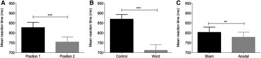
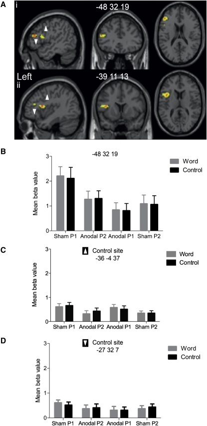
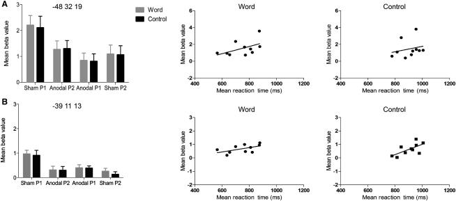
Electrophysiological studies in humans and animals suggest that noninvasive neurostimulation methods such as transcranial direct current stimulation (tDCS) can elicit long-lasting [1], polarity-dependent [2] changes in neocortical excitability. Application of tDCS can have significant and selective behavioral consequences that are associated with the cortical location of the stimulation electrodes and the task engaged during stimulation [3-8]. However, the mechanism by which tDCS affects human behavior is unclear. Recently, functional magnetic resonance imaging (fMRI) has been used to determine the spatial topography of tDCS effects [9-13], but no behavioral data were collected during stimulation. The present study is unique in this regard, in that both neural and behavioral responses were recorded using a novel combination of left frontal anodal tDCS during an overt picture-naming fMRI study. We found that tDCS had significant behavioral and regionally specific neural facilitation effects. Furthermore, faster naming responses correlated with decreased blood oxygen level-dependent (BOLD) signal in Broca's area. Our data support the importance of Broca's area within the normal naming network and as such indicate that Broca's area may be a suitable candidate site for tDCS in neurorehabilitation of anomic patients, whose brain damage spares this region.


Figures

Figure 1
Figure 1
Behavioral Effects of Anodal Transcranial Direct Current Stimulation Main effects of order, i.e., position of run during scanning session (P1 versus P2; A), cue (noise versus word; B), and stimulation (sham versus anodal tDCS [A-tDCS]; C), on naming reaction times (n = 10). Black bars are the control stimuli. Error bars indicate standard error of the mean (SEM). ∗∗∗p < 0.001, ∗∗p < 0.05.
Figure 2
Figure 2
Neural Effects of Anodal Transcranial Direct Current Stimulation (Ai and Aii) Statistical parametric maps showing the greatest reduction in BOLD (orange, peak; yellow, cluster) in inferior frontal sulcus (IFS; z score 4.98; Ai) and ventral premotor cortex (z score 4.62; Aii) as a consequence of A-tDCS. Effects of A-tDCS persist in left IFS peak (z score 4.62) and ventral premotor cortex peak (z score 3.67) when masked exclusively by order effects. (B–D) Mean beta value with SEM of each condition in IFS peak voxel (−48, 32, 19; B) illustrating decreased BOLD response associated with A-tDCS, and in left precentral gyrus (−36, −4, 37; C) and left anterior insula (−27, 32, 7; D) illustrating no effect of A-tDCS. Black bars are the control stimuli. All coordinates are in Montreal Neurological Institute space (x, y, z). See also Figures S1 and S2.
Figure 3
Figure 3
Relationship between Neural and Behavioral Effects Plots of neural effect size and correlations with naming reaction-time data for word- and noise-cued items in inferior frontal sulcus (A) and ventral premotor regions (B) affected by A-tDCS. Left panel reflects mean beta value and SEM for both significant peaks within the vicinity of left Broca's area. Scatter plots at center and right illustrate the spread and relationship between reaction time and activity in each region during sham P1 and anodal P1 conditions. Statistical analyses demonstrated a weak positive correlation in IFS between beta values and reaction time for word cues (r = 0.51, n = 10, p = 0.07, R2 = 0.26) but not noise control cues (r = 0.23, n = 10, p = 0.26). These correlations are shown in the top pair of scatter plots (left, word-cued effects; right, noise-cued effects). The bottom pair of scatter plots illustrate a significant correlation in ventral premotor cortex for word (left; r = 0.56, n = 10, p = 0.05, R2 = 0.32) and noise control cues (right; r = 0.66, n = 10, p = 0.02, R2 = 0.44, all one-tailed). Note the different scales on the y axes of the scatter plots. Anodal P1 and anodal P2 refer to the run order when A-tDCS was delivered within a scanning session. Black bars are the control stimuli.

References

    1. Fritsch B., Reis J., Martinowich K., Schambra H.M., Ji Y., Cohen L.G., Lu B. Direct current stimulation promotes BDNF-dependent synaptic plasticity: Potential implications for motor learning. Neuron. 2010;66:198–204. - PMC - PubMed
    1. Nitsche M.A., Paulus W. Excitability changes induced in the human motor cortex by weak transcranial direct current stimulation. J. Physiol. 2000;527:633–639. - PMC - PubMed
    1. de Vries M.H., Barth A.C.R., Maiworm S., Knecht S., Zwitserlood P., Flöel A. Electrical stimulation of Broca's area enhances implicit learning of an artificial grammar. J. Cogn. Neurosci. 2010;22:2427–2436. - PubMed
    1. Flöel A., Rösser N., Michka O., Knecht S., Breitenstein C. Noninvasive brain stimulation improves language learning. J. Cogn. Neurosci. 2008;20:1415–1422. - PubMed
    1. Sparing R., Dafotakis M., Meister I.G., Thirugnanasambandam N., Fink G.R. Enhancing language performance with non-invasive brain stimulation—a transcranial direct current stimulation study in healthy humans. Neuropsychologia. 2008;46:261–268. - PubMed

Publication types