. 2011 Nov;32(31):7913-23.
doi: 10.1016/j.biomaterials.2011.07.005. Epub 2011 Aug 5.

Elucidating the mechanobiology of malignant brain tumors using a brain matrix-mimetic hyaluronic acid hydrogel platform

Affiliations

Elucidating the mechanobiology of malignant brain tumors using a brain matrix-mimetic hyaluronic acid hydrogel platform

Badriprasad Ananthanarayanan et al. Biomaterials. 2011 Nov.

Abstract

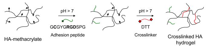
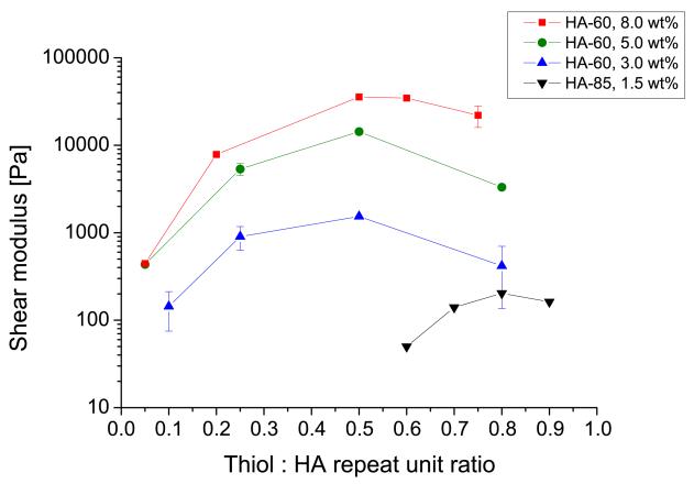
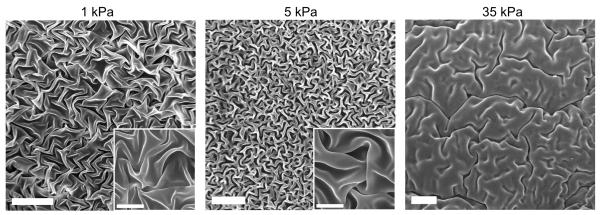
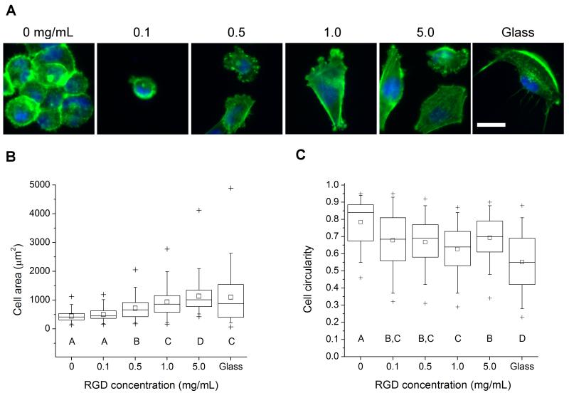
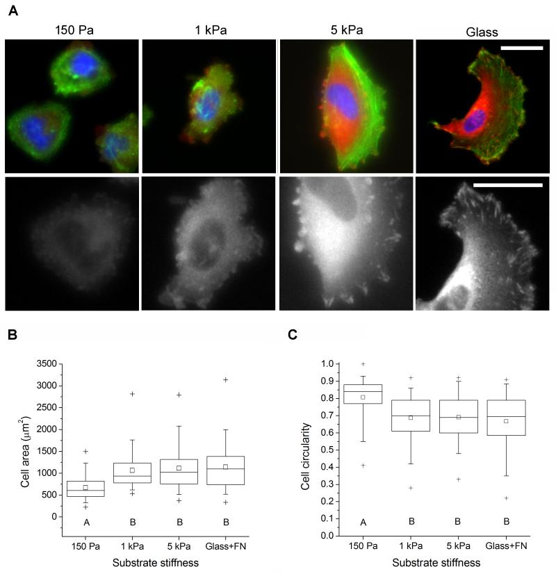
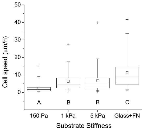
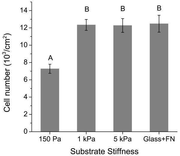
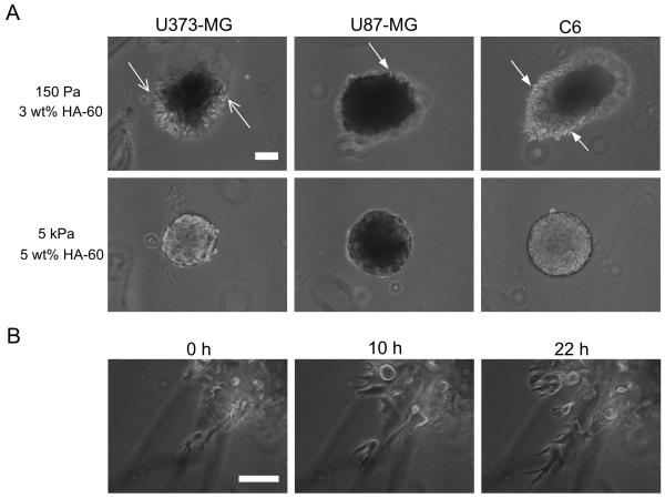
Glioblastoma multiforme (GBM) is a malignant brain tumor characterized by diffuse infiltration of single cells into the brain parenchyma, which is a process that relies in part on aberrant biochemical and biophysical interactions between tumor cells and the brain extracellular matrix (ECM). A major obstacle to understanding ECM regulation of GBM invasion is the absence of model matrix systems that recapitulate the distinct composition and physical structure of brain ECM while allowing independent control of adhesive ligand density, mechanics, and microstructure. To address this need, we synthesized brain-mimetic ECMs based on hyaluronic acid (HA) with a range of stiffnesses that encompasses normal and tumorigenic brain tissue and functionalized these materials with short Arg-Gly-Asp (RGD) peptides to facilitate cell adhesion. Scanning electron micrographs of the hydrogels revealed a dense, sheet-like microstructure with apparent nanoscale porosity similar to brain extracellular space. On flat hydrogel substrates, glioma cell spreading area and actin stress fiber assembly increased strongly with increasing density of RGD peptide. Increasing HA stiffness under constant RGD density produced similar trends and increased the speed of random motility. In a three-dimensional (3D) spheroid paradigm, glioma cells invaded HA hydrogels with morphological patterns distinct from those observed on flat surfaces or in 3D collagen-based ECMs but highly reminiscent of those seen in brain slices. This material system represents a brain-mimetic model ECM with tunable ligand density and stiffness amenable to investigations of the mechanobiological regulation of brain tumor progression.


Figures

Figure 1
Figure 1
Schematic description of HA functionalization and crosslinking. HA-methacrylate was functionalized with an RGD peptide using the Michael-type addition reaction between the methacrylate groups on the polymer and the cysteine thiol groups on the peptide. The same addition reaction with the methacrylate groups was used to induce crosslinking via reaction with dithiothreitol (DTT) to form HA hydrogels.
Figure 2
Figure 2
Mechanical characterization of HA gels. Shear elastic moduli of the DTT-crosslinked HA hydrogels were measured by oscillatory rheometry. Each curve represents HA gels containing a particular HA-methacrylate polymer (HA-60 or HA-85 with 60% and 85% degree of methacrylation, respectively) and weight fraction, at varying ratios of thiols used for crosslinking. Error bars represent standard deviation; N ≥ 2 replicates.
Figure 3
Figure 3
Scanning Electron Microscopy (SEM) imaging of dehydrated HA gels (higher magnification images in insets). The gel microstructure consists of dense, folded sheets of polymer free of fibrillar structures and does not contain micron-sized pores. The apparent density of the polymer network increases with increasing polymer weight fraction and stiffness. Scale bar = 20 μm; inset scale bar = 5 μm.
Figure 4
Figure 4
Glioma cell morphology on 35 kPa HA gels with varying surface density of RGD peptide. (A) Morphology and cytoarchitecture of cells adhered to variable-RGD density gels after 24 hr incubation, as visualized by epifluorescence imaging of F-actin (green) and nuclear DNA (blue). Scale bar = 50 μm. (B) Quantification of projected cell spreading area. (C) Quantification of cell shape, as measured by circularity (see methods). N ≥ 55 cells for each condition. Statistically distinct groups (p < 0.05) determined by Dunn’s test are marked by A, B, C, D (see methods).
Figure 5
Figure 5
Glioma cell adhesion to variable-stiffness RGD-functionalized HA gels. (A) Morphology of cells adhered to variable-stiffness gels after 24 hr incubation. Top row: immunofluorescence imaging of vinculin (orange), F-actin (green), and nuclear DNA (blue). Bottom row: Isolated vinculin signal at higher magnification. Scale bar = 50 μm. (B) Quantification of projected cell area. (C) Quantification of circularity. N ≥ 85 cells for each condition. Statistically distinct groups (p < 0.01) determined by Dunn’s test are marked by A, B (see methods).
Figure 6
Figure 6
Regulation of glioma cell motility by matrix stiffness. The plot depicts the average speed of random motility of U373-MG cells cultured on RGD-functionalized HA gels of constant peptide density and varying stiffness. N ≥ 120 cells for each condition. Statistically distinct groups (p < 0.01) determined by Dunn’s test are marked by A, B, C (see methods).
Figure 7
Figure 7
Cell number on variable-stiffness HA-RGD substrates. U373-MG cells were cultured on matrices of the specified rigidity and constant ligand density for 4 days. Cell number was then measured using the WST-1 metabolic assay. Error bars represent standard error of the mean; N = 6 replicates. Statistically distinct groups (p < 0.01) determined by Tukey’s test are marked by A, B (see methods).
Figure 8
Figure 8
3D invasion of glioma spheroids through HA-RGD hydrogels. (A) Variation of extent and patterns of invasion with cell type and matrix density. U373-MG cells dispersed and invaded as single cells (open arrows) whereas U87-MG and C6 cells retained spheroid borders with cells invading at the edges (filled arrows). No cells invaded the dense 5 kPa hydrogel. (B) Time-lapse images of U373-MG cells invading the 150 Pa hydrogel. Cells exhibited distinctly non-mesenchymal motility, with dynamically extending and branching leading processes, following by abrupt movement of the cell-body forward. Scale bar = 100 μm.

References

    1. Wen PY, Kesari S. Malignant gliomas in adults. N Engl J Med. 2008;359(5):492–507. - PubMed
    1. Nakada M, Nakada S, Demuth T, Tran NL, Hoelzinger DB, Berens ME. Molecular targets of glioma invasion. Cell Mol Life Sci. 2007;64(4):458–78. - PMC - PubMed
    1. Furnari FB, Fenton T, Bachoo RM, Mukasa A, Stommel JM, Stegh A, et al. Malignant astrocytic glioma: genetics, biology, and paths to treatment. Genes Dev. 2007;21(21):2683–710. - PubMed
    1. Pàez-Ribes M, Allen E, Hudock J, Takeda T, Okuyama H, Viñals F, et al. Antiangiogenic therapy elicits malignant progression of tumors to increased local invasion and distant metastasis. Cancer Cell. 2009;15(3):220–31. - PMC - PubMed
    1. Kunkel P, Ulbricht U, Bohlen P, Brockmann MA, Fillbrandt R, Stavrou D, et al. Inhibition of glioma angiogenesis and growth in vivo by systemic treatment with a monoclonal antibody against vascular endothelial growth factor receptor-2. Cancer Res. 2001;61(18):6624–8. - PubMed

Publication types

MeSH terms