. 2011 Sep 29;118(13):3570-8.
doi: 10.1182/blood-2011-06-359679. Epub 2011 Aug 5.

Development of thymic NK cells from double negative 1 thymocyte precursors

Affiliations

Development of thymic NK cells from double negative 1 thymocyte precursors

Claudia L Vargas et al. Blood. .

Abstract

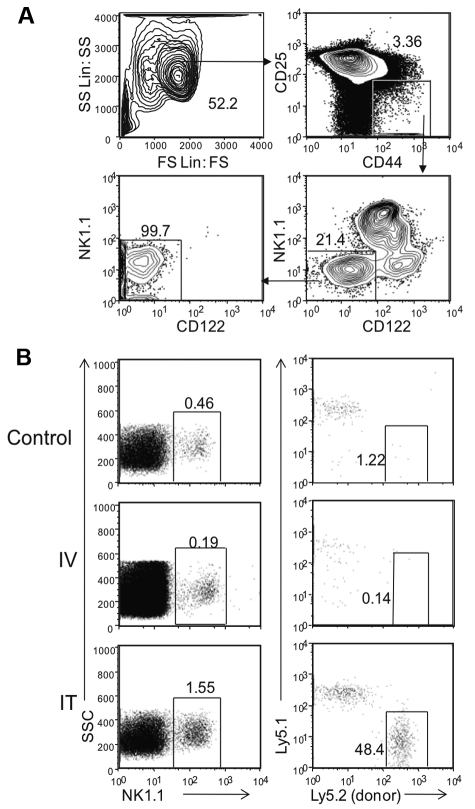
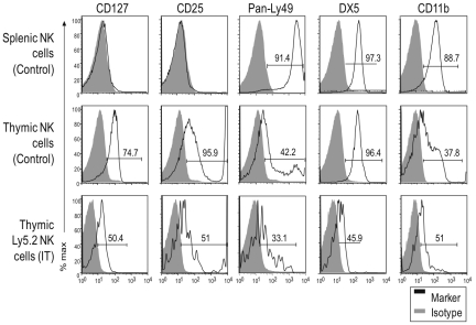
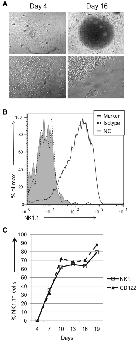
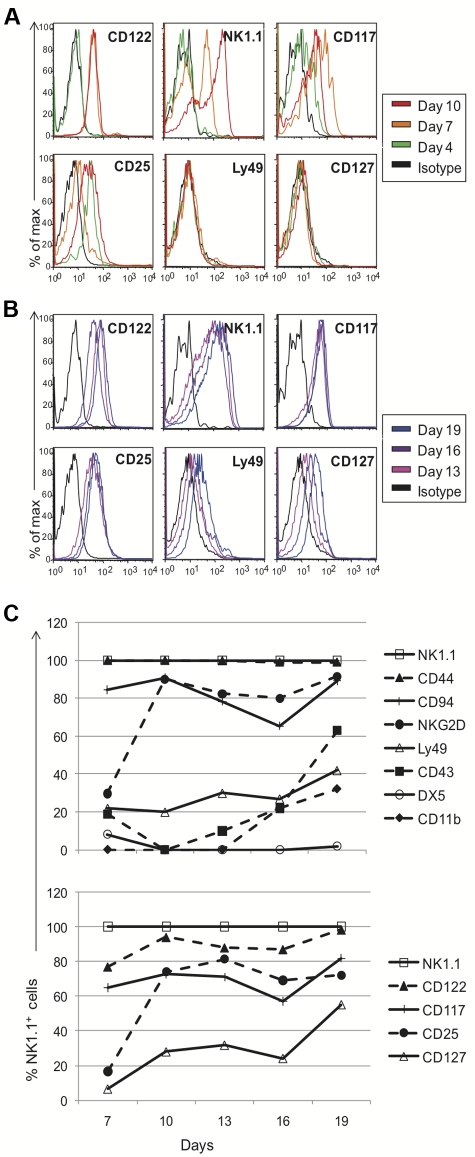
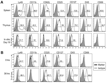
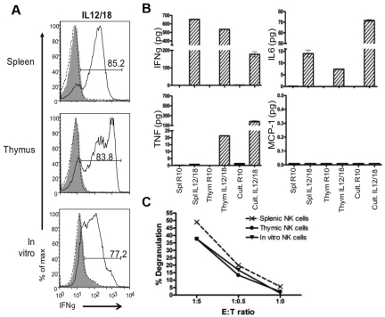
The differentiation of natural killer (NK) cells and a subpopulation of NK cells which requires an intact thymus, that is, thymic NK cells, is poorly understood. Previous in vitro studies indicate that double negative (CD4⁻CD8⁻, DN) thymocytes can develop into cells with NK cell markers, but these cells have not been well characterized. Herein, we generated and characterized NK cells differentiating from thymic DN precursors. Sorted DN1 (CD44⁺CD25⁻) CD122⁻NK1.1⁻ thymocytes from Rag1(⁻/⁻) mice were adoptively transferred into Rag1(⁻/⁻)Ly5.1 congenic mice. After intrathymic injection, donor-derived cells phenotypically resembling thymic NK cells were found. To further study their differentiation, we seeded sorted DN1 CD122⁻)NK1.1⁻ thymocytes on irradiated OP9 bone marrow stromal cells with IL-15, IL-7, Flt3L, and stem cell factor. NK1.1⁺ cells emerged after 7 days. In vitro differentiated NK cells acquired markers associated with immature bone marrow-derived NK cells, but also expressed CD127, which is typically found on thymic NK cells. Furthermore, we found that in vitro cells generated from thymic precursors secreted cytokines when stimulated and degranulated on target exposure. Together, these data indicate that functional thymic NK cells can develop from a DN1 progenitor cell population.


Figures

Figure 1
Figure 1
Donor derived DN1 CD122NK1.1 thymocytes differentiate into NK cells in the thymus. (A) Thymic DN1 CD122NK1.1 cells from Rag1−/− mice were sorted and collected for post sort analysis with the gating parameters shown. (B) Thymi from Rag1−/− Ly5.2 mice were harvested and sorted based on DN1CD122NK1.1 phenotype as shown in panel A. Cells were transferred into irradiated Rag1−/− Ly5.1 mice either intravenously or intrathymically. Irradiated littermates were used as controls. Cells were harvested from the thymus 32 days after transfer. NK1.1+ cells from the lymphocyte population were then examined for host (Ly5.1+) and donor (Ly5.2+) cells. Data are representative of 2 experiments.
Figure 2
Figure 2
DN1 CD122NK1.1 thymocytes differentiate into NK1.1+ cells with a unique phenotype. DN1 CD122NK1.1 thymocytes from Rag1−/− Ly5.2 mice were transferred into irradiated Rag1−/− Ly5.1 mice intrathymically. Donor (Ly5.2+) NK cells from IT mice were compared with NK cells from unmanipulated (nonirradiated) littermates 32 days after transfer. Gray-filled histograms represent cells stained with an isotype control and black-line histograms represent cells stained with the indicated antibody. Although the staining patterns for each marker were somewhat different, even for the same marker on different cell populations, ie, some markers were expressed on all cells while others were expressed on subsets, we used the percent of positive cells (above isotype control staining) as a convenient (although technically imprecise) means to compare and describe the staining profiles for a large number of markers. Data are representative of 2 experiments.
Figure 3
Figure 3
Thymic progenitors can differentiate into NK 1.1+ cells in vitro. Sorted DN1 CD122NK1.1 thymocytes from Rag1−/− mice were cocultured with OP9 cells and cytokines. (A) Wells were visually and microscopically examined for growth on different days. Images were acquired using a Nikon Diaphot 200 microscope (Nikon) with a Hamamatsu digital camera (Hamamatsu Photonic) and processed using MetaVue imaging software (Molecular Devices Corp). Top panels are at 4× magnification, while bottom panels are at 20× magnification. (B) Growth positive wells by visual inspection were pooled, stained, and analyzed for expression of NK1.1. For the negative controls, cells were either left unstained (gray-filled histograms) or stained using the appropriate isotype antibody (black dotted histogram). Cells were gated based on lymphocyte population by scatter parameters. Data are representative of at least 3 experiments. (C) Kinetic analysis of NK1.1 and CD122 expression on lymphocyte population, gated by scatter parameters. At various culture periods, wells were examined for the indicated markers as described in panel B. Data are representative of 3-5 experiments
Figure 4
Figure 4
Phenotypic profile of cells generated in vitro from thymic progenitors. Cells cultured in vitro were pooled on different days and assessed for marker expression via flow cytometry. Black histograms represent cells stained with appropriate isotype controls and colored-line histograms correspond to different days of analysis. Early (A) and late (B) marker acquisition is shown. Expression on gated NK1.1+ cells is shown, except for all markers on day 4 and NK1.1 expression on different days, which were gated on the lymphocyte population. (C) Developmental kinetics of sorted thymocytes as a function of days versus the percentage of NK1.1+ cells expressing the given marker. Again, as described for Figure 2, the staining patterns for each marker were somewhat different but we used the percent of positive cells as a convenient means to compare and describe the staining profiles for numerous antibodies used. Data are representative of 3-5 experiments.
Figure 5
Figure 5
In vitro developed NK cells resemble those found in the thymus and not conventional splenic NK cells. Cells from indicated tissues of Rag1−/− mice, along with in vitro differentiated NK cells were analyzed by cytometry. Gray-filled histograms represent cells stained with an isotype control and black line histograms represent cells stained with the indicated antibody. (A) In vitro generated NK cells were compared with freshly isolated splenic and thymic NK cells. Gated NK1.1+ cells are shown. (B) The phenotype of in vitro generated NK cells in culture for 19 days (top panel) was compared with that of cells that were removed from culture after 19 days and rested in LD IL15 for 36 hours (bottom panel). Gated NK1.1+ cells are shown. As described for Figure 2 and here, the staining patterns for each marker were somewhat different but we used the percentage of positive cells as a convenient means to compare and describe the staining profiles for numerous antibodies used. Data are representative of 2-5 experiments.
Figure 6
Figure 6
In vitro generated NK cells are functional. The function of in vitro differentiated NK cells grown in culture (Cult) for 19 days then rested in LD IL15 was compared with the function of Rag1−/− splenic and thymic NK cells cultured in LD IL15. Sorted NK1.1+ cells were stimulated with IL-12/IL-18 or target YAC-1 cells. (A) IFNγ production was measured by intracellular flow cytometry. For negative controls, cells were either not stained (gray-filled histograms) or not stimulated (black-dotted histograms). All histograms are gated on NK1.1+ cells. (B) Cytokines secreted were assessed by cytometric bead array. (C) CD107 degranulation on target encounter was measured by flow cytometry. Data are representative of 3 experiments.

Similar articles

Cited by

References

    1. Yokoyama WM. Chapter 17, Natural killer cells. In: Paul WE, editor. Fundamental Immunology. New York, NY: Lippincott-Raven; 2008. pp. 483–517.
    1. Loh J, Chu DT, O'Guin AK, Yokoyama WM, Virgin HW. Natural killer cells utilize both perforin and gamma interferon to regulate murine cytomegalovirus infection in the spleen and liver. J Virol. 2005;79(1):661–667. - PMC - PubMed
    1. Yokoyama WM, Kim S, French AR. The dynamic life of natural killer cells. Annu Rev Immunol. 2004;22:405–429. - PubMed
    1. Di Santo JP. Natural killer cell developmental pathways: a question of balance. Annu Rev Immunol. 2006;24:257–286. - PubMed
    1. Igarashi H, Gregory SC, Yokota T, Sakaguchi N, Kincade PW. Transcription from the RAG1 locus marks the earliest lymphocyte progenitors in bone marrow. Immunity. 2002;17(2):117–130. - PubMed

Publication types

MeSH terms