. 2011 Oct;193(20):5841-6.
doi: 10.1128/JB.00104-11. Epub 2011 Aug 5.

Processing is required for a fully functional protein P30 in Mycoplasma pneumoniae gliding and cytadherence

Affiliations

Processing is required for a fully functional protein P30 in Mycoplasma pneumoniae gliding and cytadherence

How-Yi Chang et al. J Bacteriol. 2011 Oct.

Abstract

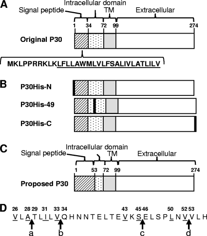
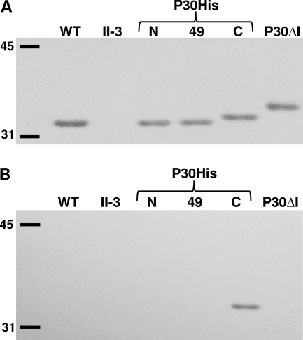
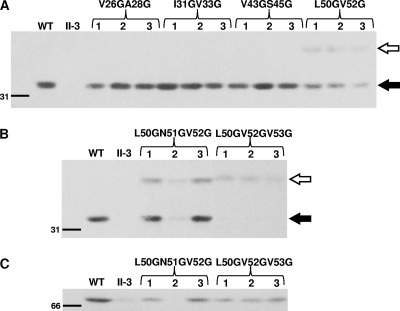
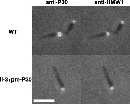
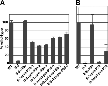
The cell wall-less prokaryote Mycoplasma pneumoniae causes bronchitis and atypical pneumonia in humans. Mycoplasma attachment to the host respiratory epithelium is required for colonization and mediated largely by a differentiated terminal organelle. P30 is an integral membrane protein located at the distal end of the terminal organelle. The P30 null mutant II-3 is unable to attach to host cells and nonmotile and has a branched cellular morphology compared to the wild type, indicating an important role for P30 in M. pneumoniae biology. P30 is predicted to have an N-terminal signal sequence, but the presence of such a motif has not been confirmed experimentally. In the current study we analyzed P30 derivatives having epitope tags engineered at various locations to demonstrate that posttranslational processing occurred in P30. Several potential cleavage sites predicted in silico were examined, and a processing-defective mutant was created to explore P30 maturation further. Our results suggested that signal peptide cleavage occurs between residues 52 and 53 to yield mature P30. The processing-defective mutant exhibited reduced gliding velocity and cytadherence, indicating that processing is required for fully functional maturation of P30. We speculate that P30 processing may trigger a conformational change in the extracellular domain or expose a binding site on the cytoplasmic domain to allow interaction with a binding partner as a part of functional maturation.


Figures

Fig. 1.
Fig. 1.
(A) Schematic of wild-type P30 based upon the originally predicted signal peptide, including the deduced amino acid sequence of the signal peptide predicted based upon its hydropathy plot (4); underlined type, predicted hydrophobic core; TM, transmembrane domain. (B) Schematic of P30His derivatives; black boxes, His6 tag. (C) Schematic of proposed P30 based upon this study. (D) P30 deduced amino acid sequence for residues 26 to 53; arrows, potential cleavage sites: a, predicted site by SignalP (NN); b, original model based upon hydropathy plot; c, predicted site by SignalP (HMM); d, predicted site by molecular mass; underlined letters, G substitutions generated. Numbers in all panels correspond to amino acid residues from N terminus.
Fig. 2.
Fig. 2.
Western immunoblot analysis of P30 from wild-type M. pneumoniae (WT), mutant II-3, the indicated P30His derivatives, and P30ΔI (4), with anti-P30 (A) or anti-poly-His (B) antibodies.
Fig. 3.
Fig. 3.
Western immunoblot analysis of representative mutant II-3 transformants with double- (A) and triple- (B and C) substitution P30 derivatives. Transformants shown in panels A and B were probed with P30-specific antibodies, and those shown in panel C with P65-specific antibodies. Three transformants are shown for each derivative. Open arrow, pre-P30; solid arrow, mature P30. Size standard is indicated on the left in kDa.
Fig. 4.
Fig. 4.
Quantitative assessment of P30 levels in wild-type M. pneumoniae (WT) and the indicated P30 derivatives by Western immunoblotting analysis. Total protein loaded: WT as indicated; all others, 40 μg. Size standard is shown on the left in kDa.
Fig. 5.
Fig. 5.
Immunofluorescence localization of P30 (left) and HMW1 (right) in wild-type M. pneumoniae (WT) and mutant II-3+pre-P30. Merged fluorescence and phase contrast images of individual cells are shown. Scale bar: 2 μm.
Fig. 6.
Fig. 6.
Quantitative analysis of HA (A) and NHBE cell colonization (B) by wild-type M. pneumoniae (WT), mutant II-3, and II-3 transformants with the indicated P30 derivatives. Values are normalized to that for wild-type M. pneumoniae. Three different transformants each for II-3+pre-P30 and II-3+tuf-pre-P30 were analyzed for HA, while one of each was examined further with NHBE cells. Bars indicate means and standard errors of the means of results from duplicate or triplicate assays.
Fig. 7.
Fig. 7.
BOXSHADE analysis of the N terminus of M. pneumoniae protein P30 and M. genitalium protein P32. Black and gray blocks, identical and similar amino acids, respectively; underlined letters, LXVV motif. The alignment of sequences was analyzed by the ClustalW2 program at http://www.ebi.ac.uk/ and box shaded by BOXSHADE 3.21 at http://www.ch.embnet.org.

Similar articles

Cited by

References

    1. Baseman J. B., et al. 1987. Identification of a 32-kilodalton protein of Mycoplasma pneumoniae associated with hemadsorption. Isr. J. Med. Sci. 23:474–479 - PubMed
    1. Biberfeld G., Biberfeld P. 1970. Ultrastructural features of Mycoplasma pneumoniae. J. Bacteriol. 102:855–861 - PMC - PubMed
    1. Biscardi S., et al. 2004. Mycoplasma pneumoniae and asthma in children. Clin. Infect. Dis. 38:1341–1346 - PubMed
    1. Chang H., Jordan J. L., Krause D. C. 2011. Domain analysis of protein P30 in Mycoplasma pneumoniae cytadherence and gliding motility. J. Bacteriol. 193:1726–1733 - PMC - PubMed
    1. Collier A. M., Clyde W. A., Jr., Denny F. W. 1971. Mycoplasma pneumoniae in hamster tracheal organ culture: immunofluorescent and electron microscopic studies. Proc. Soc. Exp. Biol. Med. 136:569–573 - PubMed

Publication types

MeSH terms

LinkOut - more resources