

doi: 10.1107/S1744309111024249.
Epub 2011 Jul 26.
Structure of filamin A immunoglobulin-like repeat 10 from Homo sapiens
Affiliations
- PMID: 21821884
- PMCID: PMC3151117
- DOI: 10.1107/S1744309111024249
Item in Clipboard
Structure of filamin A immunoglobulin-like repeat 10 from Homo sapiens
Acta Crystallogr Sect F Struct Biol Cryst Commun.
.
Abstract
Filamin A (FlnA) plays a critical role in cytoskeletal organization, cell motility and cellular signaling. FlnA utilizes different binding sites on a series of 24 immunoglobulin-like domains (Ig repeats) to interact with diverse cytosolic proteins and with cytoplasmic portions of membrane proteins. Mutations in a specific domain, Ig10 (FlnA-Ig10), are correlated with two severe forms of the otopalatodigital syndrome spectrum disorders Melnick-Needles syndrome and frontometaphyseal dysplasia. The crystal structure of FlnA-Ig10 determined at 2.44 Å resolution provides insight into the perturbations caused by these mutations.
Figures

Crystal structure of human FlnA-Ig10. (a) Ribbon diagram of FlnA-Ig10 colored as a rainbow from the N-terminus (blue) to the C-terminus (red). β-Strands A–G and loops are labeled. (b) A disulfide bond (yellow) between two symmetry-related copies of FlnA-Ig10: chain A from the asymmetric unit (magenta) and chain B of a symmetry mate (x + 1/2, −y + 1/2, −z) (cyan). (c) FlnA-Ig10 (magenta) coordinates an acetate ion using a water-mediated hydrogen-bond network utilizing the Tyr1229 hydroxyl, the Leu1224 backbone amide and a water molecule (red sphere) hydrogen bonded to the Ile1222 backbone carbonyl. (d) Cys1198 is covalently modified by a β-mercaptoethanol adduct. The β-mercaptoethanol-modified Cys1198 lies on the solvent-exposed surface of β-sheet C. 2F
o − F
c density (dark blue) is contoured at 1.2σ in (b)–(d).
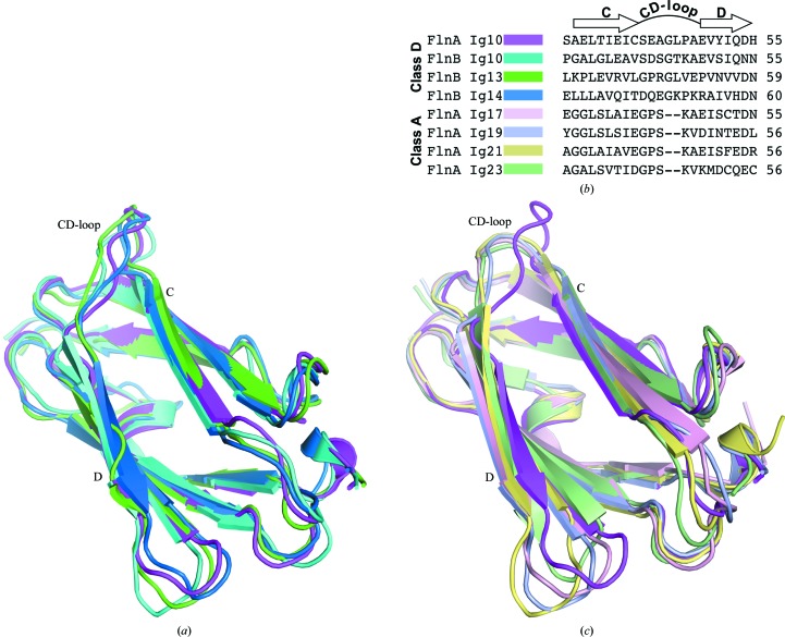
Comparison of FlnA-Ig10 to existing structures of filamin Ig repeats. (a) Overlay of structures for the class D filamin Ig repeats FlnA-Ig10 (magenta), FlnB-Ig10 (cyan; PDB entry 2dia ), FlnB-Ig13 (green; PDB entry 2dj4 ) and FlnB-Ig14 (blue; PDB entry 2e9j ) (T. Tomizawa, S. Koshiba, S. Watanabe, T. Harada, T. Kigawa & S. Yokoyama, unpublished work). (b) Sequence alignment of the CD-loop region of class D filamin Ig repeats (FlnA-Ig10, FlnB-Ig10, FlnB-Ig13 and FlnA-Ig14) and class A filamin Ig repeats (FlnA-Ig17, FlnA-Ig19, FlnA-Ig21 and FlnA-Ig23). (c) Overlay of the FlnA-Ig10 (magenta) structure with class A filamin Ig repeats FlnA-Ig17 (light pink; PDB entry 2bp3 ; Nakamura et al., 2006 ▶), FlnA-Ig19 (light blue; PDB entry 2j3s ; Lad et al., 2008 ▶), FlnA-Ig21 (light yellow; PDB entry 3isw ; Smith et al., 2010 ▶) and FlnA-Ig23 (light green; PDB entry 2k3t ; Nakamura et al., 2009 ▶).
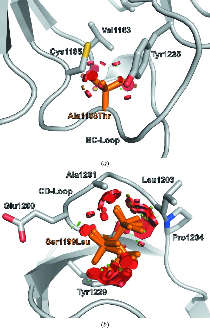
Locations of mutations correlated with MNS. (a) Ala1188 of the BC-loop is not solvent-accessible and abuts the FlnA-Ig10 hydrophobic core. Backbone-dependent threonine rotamers for the A1188T mutation result in clashes (red disks) with neighboring residues (gray sticks). (b) Ser1199 within the CD-loop displays low side-chain solvent accessibility and all backbone-dependent leucine rotamers for the S1199L mutation lead to steric clashes (red disks) with surrounding residues (gray sticks).
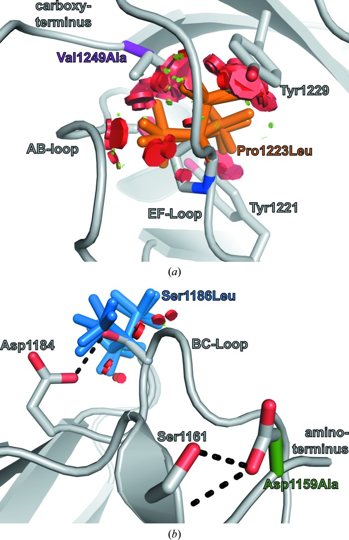
Two distinct FlnA-Ig10 regions harbor mutations correlated with FMD. (a) All preferred leucine rotamers for mutation P1223L (orange) result in significant steric clashes (red discs) with surrounding residues (gray sticks). Although mutation V1249A (purple) would not result in steric clashes, it may lead to a hydrophobic packing defect. (b) Mutations D1159A (green) and S1186L (blue) occur in surface-exposed residues. Each mutation can be accommodated by preferred rotamers without significant steric clashes (red disks); however, each mutation would preclude the formation of side-chain-mediated hydrogen bonds (black dashes).
References
-
- Adams, P. D. et al. (2010). Acta Cryst. D66, 213–221.
-
- Bork, P., Holm, L. & Sander, C. (1994). J. Mol. Biol. 242, 309–320. - PubMed
-
- DeLano, W. L. (2002). PyMOL http://www.pymol.org.
Publication types
MeSH terms
Substances
Associated data
- Actions
Grants and funding
LinkOut - more resources
Full Text Sources
Miscellaneous
