. 2011 Aug 1;67(Pt 8):947-52.
doi: 10.1107/S1744309111019300. Epub 2011 Jul 26.

Purification, crystallization and preliminary X-ray diffraction analysis of the human mismatch repair protein MutSβ

Affiliations

Purification, crystallization and preliminary X-ray diffraction analysis of the human mismatch repair protein MutSβ

Quincy Tseng et al. Acta Crystallogr Sect F Struct Biol Cryst Commun. .

Abstract

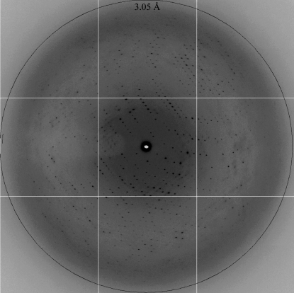
MutSβ is a eukaryotic mismatch repair protein that preferentially targets extrahelical unpaired nucleotides and shares partial functional redundancy with MutSα (MSH2-MSH6). Although mismatch recognition by MutSα has been shown to involve a conserved Phe-X-Glu motif, little is known about the lesion-binding mechanism of MutSβ. Combined MSH3/MSH6 deficiency triggers a strong predisposition to cancer in mice and defects in msh2 and msh6 account for roughly half of hereditary nonpolyposis colorectal cancer mutations. These three MutS homologs are also believed to play a role in trinucleotide repeat instability, which is a hallmark of many neurodegenerative disorders. The baculovirus overexpression and purification of recombinant human MutSβ and three truncation mutants are presented here. Binding assays with heteroduplex DNA were carried out for biochemical characterization. Crystallization and preliminary X-ray diffraction analysis of the protein bound to a heteroduplex DNA substrate are also reported.


Figures

Figure 1
Figure 1
Purification of MutSβ and its deletion mutants. (a) Representative chromatogram (shown for MutSβΔ162) of the final purification step (Mono Q column) for MutSβ and its deletion mutants. Absorbance at 280 nm versus elution volume is shown in blue; percentage elution buffer versus elution volume is shown in red. (b) A 12% SDS–PAGE of purified human MutSβ and its deletion mutants. Lane 1 shows molecular-weight standards, with weights (in kDa) indicated on the left. Lanes 2, 3, 4 and 5 show full-length MutSβ (FL), MutSβΔ162, MutSβΔ175 and MutSβΔ223. The locations of both MSH2 and MSH3 subunits are indicated by labelled arrows.
Figure 3
Figure 3
Binding of MutSβ and MutSβΔ162 to a 41 bp heteroduplex containing a CA insertion–deletion loop. (a) Binding of MutSβ (circles) and MutSβΔ162 (squares) to a 41 bp heteroduplex (closed symbols) or control homoduplex (open symbols) was assessed by SPRS (see §2.3). Mass response units at saturation (∼400 s) are plotted as a function of protein concentration. Data were fitted by nonlinear least-squares regression to a rectangular hyperbola, yielding K d values of 4 ± 1 nM for the wild-type protein and 5 ± 2 nM for MutSβΔ162 for heteroduplex binding. Binding to homoduplex DNA was not saturable up to 100 nM protein concentration. Dissociation of MutSβ (b) or MutSβΔ162 (c) from 41 bp heteroduplex (solid lines) and homoduplex (dotted lines) DNA substrates upon challenge (arrow) with 1 mM ATP was assessed by SPRS.
Figure 4
Figure 4
Human MutSβ crystals. (a) Tetragonal form 1 crystal with approximate dimensions of 200 × 200 × 200 µm. (b) Monoclinic form 2 crystals of dimensions 200 × 80 × 40 µm.
Figure 2
Figure 2
SDS–PAGE of limited proteolysis of human MutSβ by subtilisin. The leftmost lane shows molecular-weight standards, with weights (in kDa) indicated on the left. Digestion times (min) are shown at the top. An MSH3 fragment was detected starting 2 min after digestion. The locations of full-length (FL) MSH3, the MSH3 proteolysis fragment and MSH2 are indicated by labelled arrows.
Figure 5
Figure 5
A diffraction pattern of crystal form 2 collected on beamline 12.3.1 at the Advanced Light Source. The resolution ring corresponds to 3.05 Å.

References

    1. Acharya, S., Wilson, T., Gradia, S., Kane, M. F., Guerrette, S., Marsischky, G. T., Kolodner, R. & Fishel, R. (1996). Proc. Natl Acad. Sci. USA, 93, 13629–13634. - PMC - PubMed
    1. Berndt, S. I., Platz, E. A., Fallin, M. D., Thuita, L. W., Hoffman, S. C. & Helzlsouer, K. J. (2007). Int. J. Cancer, 120, 1548–1554. - PubMed
    1. Blackwell, L. J., Wang, S. & Modrich, P. (2001). J. Biol. Chem. 276, 33233–33240. - PubMed
    1. Broek, W. J. van den, Nelen, M. R., Wansink, D. G., Coerwinkel, M. M., te Riele, H., Groenen, P. J. & Wieringa, B. (2002). Hum. Mol. Genet. 11, 191–198. - PubMed
    1. Dowen, J. M., Putnam, C. D. & Kolodner, R. D. (2010). Mol. Cell. Biol. 30, 3321–3328. - PMC - PubMed

Publication types

Substances