. 2011;34(3):268-80.
doi: 10.1159/000330187. Epub 2011 Aug 4.

Glomerular expression of kidney injury molecule-1 and podocytopenia in diabetic glomerulopathy

Affiliations

Glomerular expression of kidney injury molecule-1 and podocytopenia in diabetic glomerulopathy

Xueying Zhao et al. Am J Nephrol. 2011.

Abstract

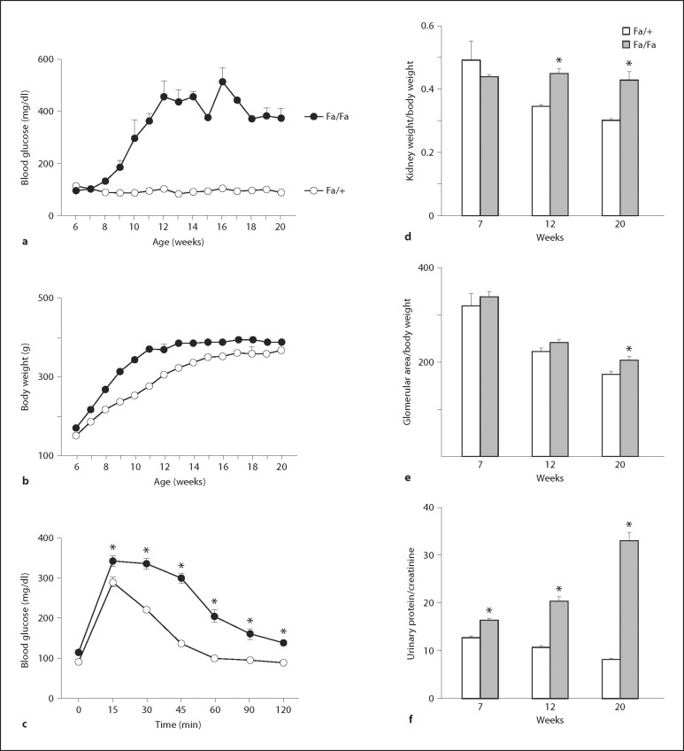
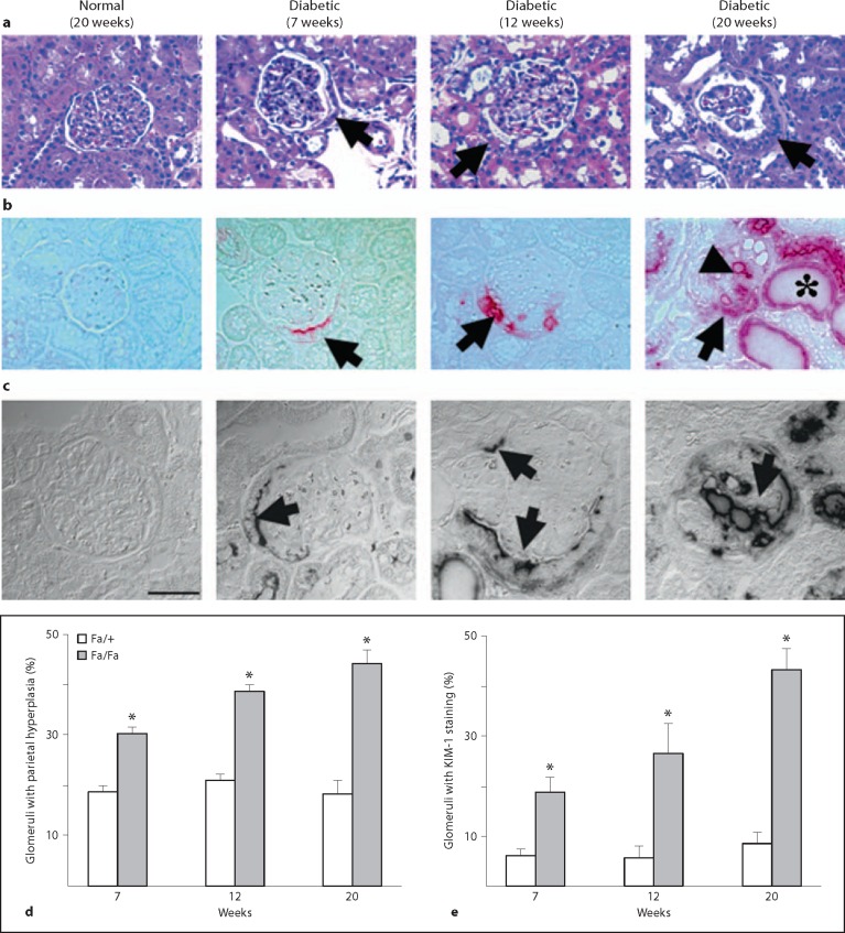
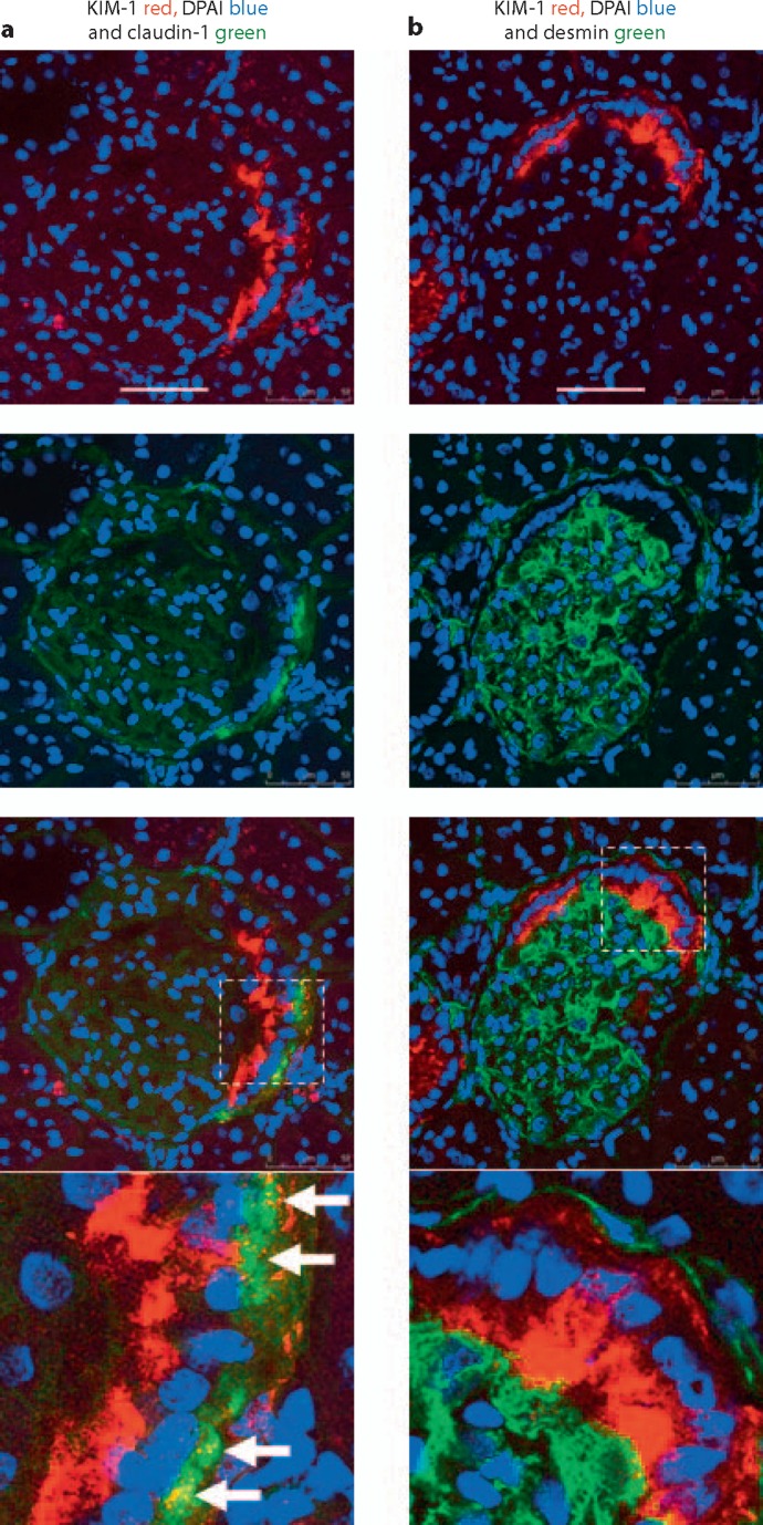
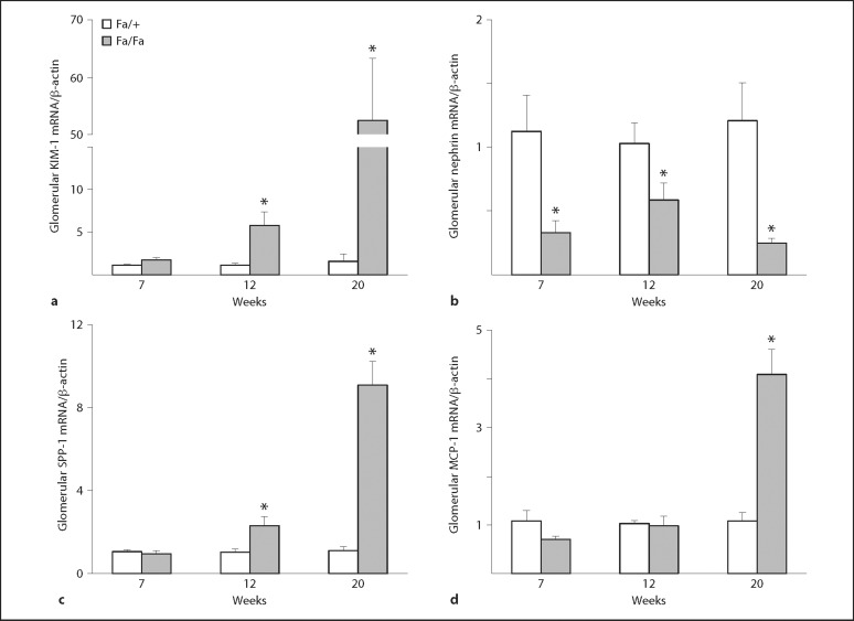
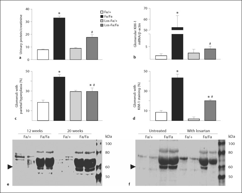
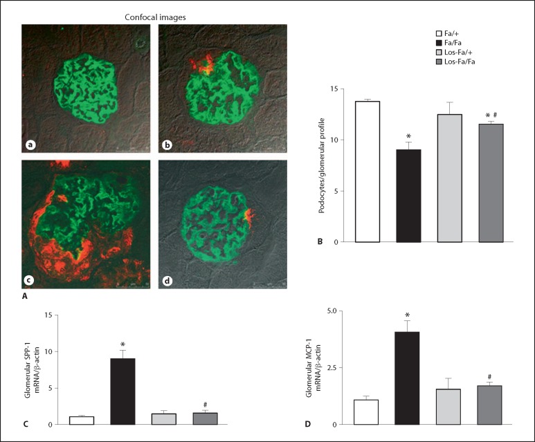
Background/aims: Studies have shown that kidney injury molecule-1 (KIM-1) is upregulated in damaged renal proximal tubules. In this study, we examined KIM-1 expression in glomerular epithelial cells in diabetic glomerulopathy.

Methods: Renal histology, immunostaining and Western blot for protein level, and real-time PCR for mRNA expression of KIM-1 and podocyte markers were evaluated in untreated or losartan-treated Zucker lean (Fa/+) and Zucker diabetic fatty (Fa/Fa) rats.

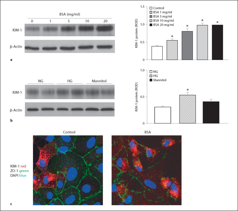
Results: The diabetic rats showed an increased glomerular expression of KIM-1. KIM-1 staining was localized primarily in the hyperplastic parietal epithelium of Bowman's capsule in the early stages of diabetes with subsequent increase in KIM-1-positive cells in the glomerular tuft in the more advanced stages. The increase in glomerular KIM-1 was associated with a decrease in podocytes in Fa/Fa rats. Antiproteinuric treatment with losartan attenuated podocytopenia and decreased renal expression of KIM-1 in treated diabetic rats. In an in vitro study, albumin overload increased KIM-1 protein in the primary cultures of rat glomerular epithelial cells.

Conclusion: These results show that glomerular KIM-1 expression was increased, in proportion to the extent of proteinuria and podocytopenia in the diabetic animals, supporting that KIM-1 could be used as a potential biomarker for glomerular injury in proteinuric kidney disease.


Figures

Fig. 1
Fig. 1
Metabolic values in the Zucker lean (Fa/+) and Zucker diabetic fatty (Fa/Fa) animals. Nonfasting blood glucose levels (a) and body weight (b) were measured at weekly intervals. c Blood glucose responses to an intraperitoneal glucose challenge were determined in 7-week-old Fa/+ and Fa/Fa rats. d and e present the ratios of left kidney weight (d) and glomerular surface area (e) to total body weight. f The urinary protein to creatinine ratio was obtained by dividing the urinary protein concentration by the urine creatinine concentration, both expressed in mg/dl. Values are mean ± SEM. n = 5–10 animals/group. * p < 0.05 vs. Fa/+ littermates.
Fig. 2
Fig. 2
Increased parietal epithelial hyperplasia and KIM-1-positive staining in the glomeruli of diabetic animals. a Representative HE staining images show increased cuboidal (hyperplastic) PECs (arrow) of Bowman's capsule in Zucker diabetic fatty rats compared to Zucker lean normal controls. Note the presence of a visceral-parietal epithelial bridge in the glomerulus of diabetic rats (12 weeks). b KIM-1 red-positive staining in the hyperplastic parietal epithelium (arrow), glomerular tuft (arrow head), and proximal tubular epithelium (*) in the diabetic animals. Original magnification ×400. c Confocal DIC images of KIM-1 (arrow) in the glomeruli of Zucker diabetic kidneys. No KIM-1 staining in the glomerulus of normal Fa/+ rats. In 7-week-old diabetic Fa/Fa rats, KIM-1-specific staining (black) was seen in the apical membrane of hyperplastic PECs. Increased KIM-1 staining was also observed in the glomerular tuft in connection with KIM-1-positive parietal epithelium in the kidney section of 20-week-old Fa/Fa rats. DIC = Differential interference contrast. Scale bar = 50 μm. d, e Quantitative analysis of the percentage of glomeruli with parietal hyperplasia (d) or KIM-1-positive staining (e) in the Zucker lean (Fa/+) and diabetic fatty (Fa/Fa) rats at the ages of 7–20 weeks. Values are mean ± SEM. n = 5–6 animals/group. * p < 0.05 vs. Fa/+ littermates.
Fig. 3
Fig. 3
Representative confocal images of triple staining for nephrin (green), WT-1 (red) and KIM-1 (blue) in the glomeruli of Zucker animals. a Normal glomerulus presents nephrin- and WT-1-positive but KIM-1-negative staining. b–d Decreased nephrin and WT-1 in association with an increase in KIM-1 blue staining in the glomeruli of Fa/Fa diabetic animals at the age of 12 (b), 20 (c) and 28 (d) weeks. Scale bars = 50 μm. Colors refer to the online version only.
Fig. 4
Fig. 4
Representative confocal images of double staining for KIM-1 with claudin-1 (PEC marker) or KIM-1 with desmin (podocyte injury marker) in the glomeruli of 20-week-old Fa/Fa rats. a Colocalization of KIM-1 (red) and claudin-1 (green) was seen in the basolateral membrane of PECs (white arrows). b There was no significant overlap between KIM-1 (red) and desmin (green) in the parietal epithelium of Bowman's capsule. Scale bars = 50 μm. Colors refer to the online version only.
Fig. 5
Fig. 5
mRNA levels of glomerular KIM-1, nephrin, SPP-1 and MCP-1. Glomerular KIM-1 (a), nephrin (b), SPP-1 (c) and MCP-1 (d) mRNA levels were evaluated by TaqMan real-time PCR. Values are mean ± SEM. n = 5–7 animals/group. * p < 0.05 vs. Fa/+ littermates.
Fig. 6
Fig. 6
Effects of losartan treatment on KIM-1 expression and urinary protein excretion. 12-week-old Zucker rats were treated with losartan (Los-Fa/+ and Los-Fa/Fa) for 8 weeks and their urinary protein to creatinine ratio (a), glomerular KIM-1 mRNA (b), percentage of glomeruli with parietal hyperplasia (c), and percentage of glomeruli with KIM-1-positive staining (d) were compared with those in untreated Fa/+ or Fa/Fa groups. e Representative Western blot image shows a progressive increase in urinary KIM-1 protein (∼60 kDa, arrowhead) in 12- to 20-week-old Fa/Fa diabetic rats compared to the Fa/+ normal controls. f Urinary excretion of KIM-1 in the diabetic rats was attenuated markedly by losartan treatment. Values are mean ± SEM. n = 4–7 animals/group. * p < 0.05 vs. normal Fa/+ group; # p < 0.05 vs. untreated Fa/Fa group.
Fig. 7
Fig. 7
Effects of losartan on expression of nephrin and inflammatory markers. A Representative confocal images of double staining for nephrin (green) and KIM-1 (red) in the glomeruli of 20-week-old Fa/+ normal controls (a), 12-week-old Fa/Fa (b), untreated 20-week-old Fa/Fa (c) and losartan-treated Fa/Fa (d) rats. Colors refer to the online version only. B Bar graph presents the number of podocytes per glomerular section in 20-week-old untreated (Fa/+ and Fa/Fa) and losartan-treated (Los-Fa/+ and Los-Fa/Fa) animals. C, D Impact of losartan on mRNA levels of SPP-1 and MCP-1. The diabetes-related increase in renal expression of SPP-1 (C) and MCP-1 (D) genes was prevented by chronic treatment with losartan. Values are mean ± SEM. n = 4–7 animals/group. * p < 0.05 vs. Fa/+ normal control; # p < 0.05 vs. untreated Fa/Fa diabetic group.
Fig. 8
Fig. 8
KIM-1 protein expression in primary cultures of rat glomerular epithelial cells in response to bovine serum albumin (BSA) and high glucose (HG) stimulation. a Western blot analysis shows that BSA (1–20 mg/ml) treatment resulted in a dose-dependent increase in KIM-1 protein in glomerular epithelial cells in primary culture. b KIM-1 protein was modestly increased following 72-hour incubation with high glucose. 4–6 epithelial cultures were treated for each condition; * p < 0.05 vs. untreated control group. c Confocal images show that primary cultured glomerular epithelial cells express KIM-1, which was markedly increased in the presence of 10 mg/ml albumin. Expression of KIM-1 was accompanied by disruption of normal characteristic ZO-1 staining at cell-cell contact of epithelial monolayer. Scale bars = 25 μm. NG = Normal glucose.

References

    1. de Zeeuw D, Remuzzi G, Parving HH, et al. Albuminuria, a therapeutic target for cardiovascular protection in type 2 diabetic patients with nephropathy. Circulation. 2004;110:921–927. - PubMed
    1. Siu B, Saha J, Smoyer WE, et al. Reduction in podocyte density as a pathologic feature in early diabetic nephropathy in rodents: prevention by lipoic acid treatment. BMC Nephrol. 2006;7:6. - PMC - PubMed
    1. Stitt-Cavanagh E, MacLeod L, Kennedy C. The podocyte in diabetic kidney disease. Sci World J. 2009;9:1127–1139. - PMC - PubMed
    1. Hoshi S, Shu Y, Yoshida F, et al. Podocyte injury promotes progressive nephropathy in Zucker diabetic fatty rats. Lab Invest. 2002;82:25–35. - PubMed
    1. Ohse T, Vaughan MR, Kopp JB, et al. De novo expression of podocyte proteins in parietal epithelial cells during experimental glomerular disease. Am J Physiol Renal Physiol. 2010;298:F702–F711. - PMC - PubMed

Publication types

Substances