. 2011 Aug 15;10(16):2806-15.
doi: 10.4161/cc.10.16.17012. Epub 2011 Aug 15.

Characterization of the cell of origin for small cell lung cancer

Affiliations

Characterization of the cell of origin for small cell lung cancer

Kwon-Sik Park et al. Cell Cycle. .

Abstract

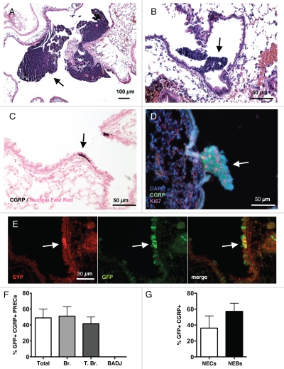
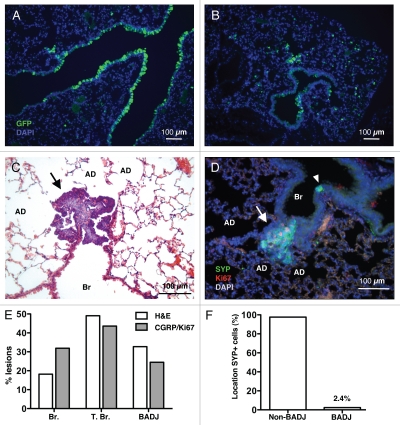
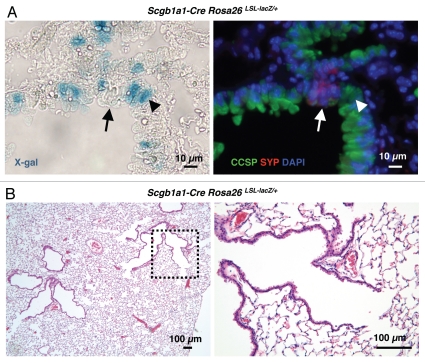
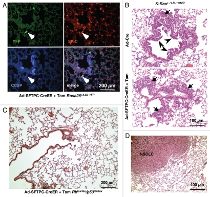
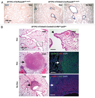
Small cell lung carcinoma (SCLC) is a neuroendocrine subtype of lung cancer that affects more than 200,000 people worldwide every year with a very high mortality rate. Here, we used a mouse genetics approach to characterize the cell of origin for SCLC; in this mouse model, tumors are initiated by the deletion of the Rb and p53 tumor suppressor genes in the lung epithelium of adult mice. We found that mouse SCLCs often arise in the lung epithelium, where neuroendocrine cells are located, and that the majority of early lesions were composed of proliferating neuroendocrine cells. In addition, mice in which Rb and p53 are deleted in a variety of non-neuroendocrine lung epithelial cells did not develop SCLC. These data indicate that SCLC likely arises from neuroendocrine cells in the lung.


Figures

Figure 1
Figure 1
Induction of neuroendocrine lesions and SCLC in the lungs of mice. Hematoxylin and Eosin (H&E) staining of lungs sections from Rb/p53 mutant mice infected with Ad-Cre and aged for 9–12 mo (A) or 3–6 mo (B). Arrows indicate large tumors and small lesions, respectively. (C) Immunostaining for the neuroendocrine marker CGRP (dark brown, arrow) in a section from wild-type mouse lungs counterstained with Nuclear Fast Red. (D) Immunofluorescence analysis of CGRP (green) and Ki67 (red) expression in the lungs of Rb/p53 mutant mice infected with Ad-Cre and aged for 3 mo. DAPI (blue) stains DNA. (E) Immunofluorescence analysis of Synaptophysin (SYP) and GFP expression on a lung section from a mouse infected with Ad-GFP. (F) Quantification of CGRP/GFP double-positive cells in sections from Rb/p53 mutant lungs. (G) Quantification of GFP expression in isolated neuroendocrine cells (NECs) and neuroendocrine bodies (NEBs, defined by clusters of NECs). All pictures are representative of multiple mice. Data for (F and G) come from the analysis of 55 pulmonary neuroendocrine cells or cell clusters from 16 mutant mice.
Figure 2
Figure 2
Early neuroendocrine lesions in various locations along the lung epithelium. Immunostaining for GFP on lung sections from mice infected with Ad-GFP three days before analysis shows that cells lining airways (A) and in the alveolar space (B) express GFP. (C) H&E staining of lung sections from one representative Rb/p53 mutant mouse infected with Ad-Cre and aged for 3 mo. AD, alveolar duct; Br, bronchioles. A typical SCLC lesion with scant cytoplasm and hyperchromatic nuclei (arrow) can be identified at the BADJ. (D) Immunostaining for SYP and Ki67 identifies actively dividing neuroendocrine lesions (arrow). A quiescent NEB is present on the same section (arrowhead). (E) Quantification of the localization of lesions identified by histological criteria in H&E sections (as in C, n = 55 from 16 mice, white bars) and by double staining for SYP and Ki67 (as in D, n = 94 from 25 mice, gray bars). T. Br., terminal bronchioles. (F) Quantification of the localization of SYP+ cells in sections from normal lungs (n = 586 neuroendocrine bodies). Any SYP+ cell within ten cells of the BADJ was counted in the BADJ group.
Figure 3
Figure 3
Constitutive deletion of Rb and p53 in CCSP-expressing cells. (A) Representative X-gal staining of lung sections from an Scgb1a1-Cre Rosa26R mouse (left). The section was also immunostained for CCSP and SYP expression to identify Clara cells (arrowhead) and pulmonary neuroendocrine cells (arrow), respectively (right). (B) H&E staining of a representative lung section from an Scgb1a1-Cre Rb/p53 mutant mouse. The dotted area in the left part is magnified in the right part. No tumors or small lesions are visible.
Figure 4
Figure 4
Deletion of Rb and p53 in SP-C-expressing adult cells using adenovirus. (A) Representative immunofluorescence staining of lung sections from a Rosa26+/LSL-YFP reporter mouse infected with Ad-SFTPC-CreER following activation of Cre by Tam. The arrow points to a cell that is YFP+ and SP-C+ but CCSP (B) Development of bronchiolar hyperplasia (arrowheads) and atypical alveolar hyperplasia (AAH) (arrows) 6–8 weeks after infection of K-Ras+/LSL-G12D with Ad-Cre (Top). The same mice infected with Ad-SFTPC-CreER and injected with Tam only develop AAH (Bottom). (C) H&E staining of a representative lung section from an Rb/p53 mutant mouse infected with Ad-SFTPC-CreER and injected with Tam and aged over 9 mo. The image is representative of multiple mice. (D) Development of NSCLC in an Rb/p53 mutant mouse infected with Ad-SFTPC-CreER and injected with Tam and aged over 9 mo.
Figure 5
Figure 5
Induced deletion of Rb and p53 in SP-C-expressing cells. (A) X-gal-stained lung sections from Sftpc-rtTA/tetO-Cre/Rosa26R mice treated with doxycycline or vehicle (Dox or No Dox, respectively). The lungs of Sftpc-rtTA/Rosa26R mice (Dox, far left) were used as a negative control. (B) Representative H&E staining and immunostaining for proSP-C and Ki67 on lung sections from Sftpc-rtTA/tetO-Cre/Rblox/lox/p53lox/lox mice treated with doxycycline or vehicle. The dotted area in the top left part is highlighted in the right part. The arrowhead in the right part indicates normal pulmonary neuroendocrine cell. Tumor cells (arrows) in both Dox and No Dox groups stained positive for proSP-C, a maker of adenocarcinoma as well as alveolar type II cells and Ki67, a marker of proliferation.

Comment in

References

    1. Beasley MB, Brambilla E, Travis WD. The 2004 World Health Organization classification of lung tumors. Semin Roentgenol. 2005;40:90–97. doi: 10.1053/j.ro.2005.01.001. - DOI - PubMed
    1. Travis WD, Travis LB, Devesa SS. Lung cancer. Cancer. 1995;75:191–202. doi: 10.1002/1097-0142(19950101)75:1+<191::AID-CNCR2820751307>3.0.CO;2-Y. - DOI - PubMed
    1. Worden FP, Kalemkerian GP. Therapeutic advances in small cell lung cancer. Expert Opin Investig Drugs. 2000;9:565–579. doi: 10.1517/13543784.9.3.565. - DOI - PubMed
    1. Socinski MA, Bogart JA. Limited-stage small-cell lung cancer: the current status of combined-modality therapy. J Clin Oncol. 2007;25:4137–4145. doi: 10.1200/JCO.2007.11.5303. - DOI - PubMed
    1. Adelstein DJ, Tomashefski J, Jr, Snow NJ, Horrigan TP, Hines JD. Mixed small cell and non-small cell lung cancer. Chest. 1986;89:699–704. doi: 10.1378/chest.89.5.699. - DOI - PubMed

Publication types

Substances