. 2011 Aug 5;30(19):3994-4005.
doi: 10.1038/emboj.2011.272.

Nucleophosmin deposition during mRNA 3' end processing influences poly(A) tail length

Affiliations

Nucleophosmin deposition during mRNA 3' end processing influences poly(A) tail length

Fumihiko Sagawa et al. EMBO J. .

Abstract

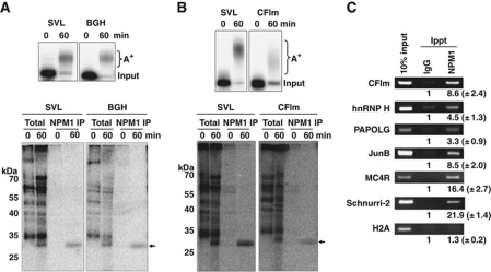
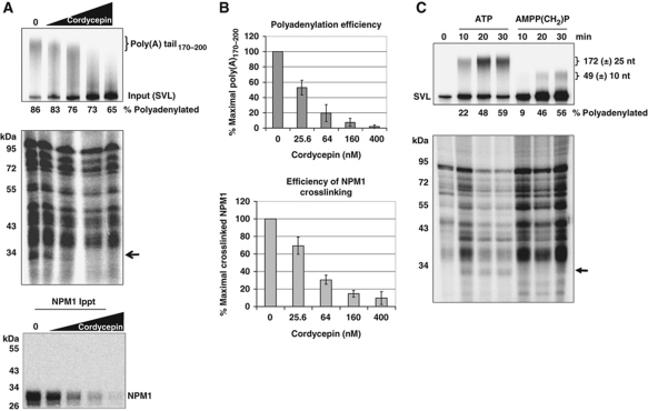
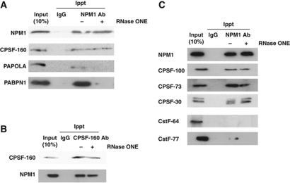
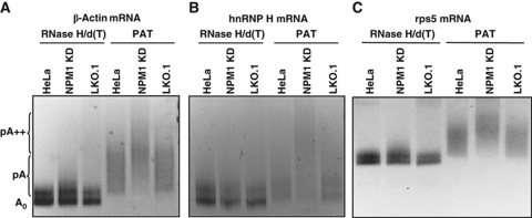
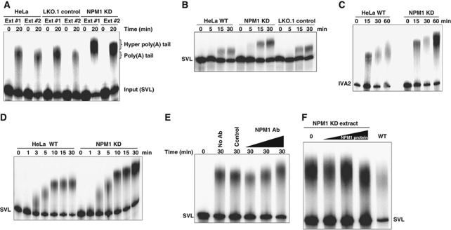
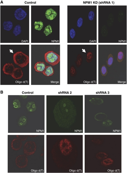
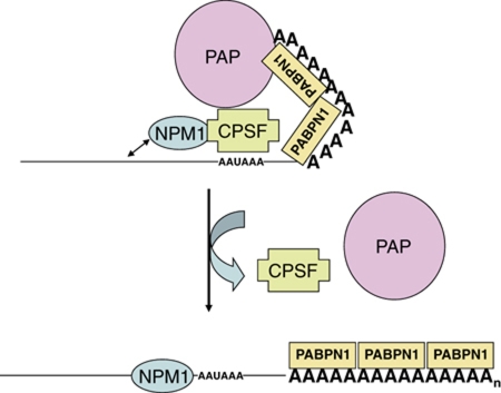
During polyadenylation, the multi-functional protein nucleophosmin (NPM1) is deposited onto all cellular mRNAs analysed to date. Premature termination of poly(A) tail synthesis in the presence of cordycepin abrogates deposition of the protein onto the mRNA, indicating natural termination of poly(A) addition is required for NPM1 binding. NPM1 appears to be a bona fide member of the complex involved in 3' end processing as it is associated with the AAUAAA-binding CPSF factor and can be co-immunoprecipitated with other polyadenylation factors. Furthermore, reduction in the levels of NPM1 results in hyperadenylation of mRNAs, consistent with alterations in poly(A) tail chain termination. Finally, knockdown of NPM1 results in retention of poly(A)(+) RNAs in the cell nucleus, indicating that NPM1 influences mRNA export. Collectively, these data suggest that NPM1 has an important role in poly(A) tail length determination and may help network 3' end processing with other aspects of nuclear mRNA maturation.


Conflict of interest statement

The authors declare that they have no conflict of interest.

Figures

Figure 1
Figure 1
Nucleophosmin is deposited on cellular poly(A)+ mRNAs in both HeLa cells and an in vitro polyadenylation system. (A, B) Radiolabelled RNAs containing polyadenylation signals from the SV40 late (SVL), bovine growth hormone (BGH) or the 68-kDa component of cleavage factor Im (CFIm) were incubated with HeLa nuclear extract in an in vitro polyadenylation assay for the times indicated. RNA products were analysed on a 5% acrylamide gel containing urea (top panel). A+ indicates the migration of the polyadenylated product RNAs, while ‘input’ indicates the size of the RNA substrate. In the lower panel, in parallel experiments in the cell-free polyadenylation system radioactive RNAs were crosslinked by UV light to closely associated proteins, treated with RNase and total crosslinked proteins were analysed by SDS–PAGE (total lanes) or immunoprecipitated using an anti-NPM1 antisera before electrophoresis (NPM1 IP lanes). The arrow on the right indicates the migration of the NPM1 crosslinked product while molecular weight markers are indicated on the left. (C) HeLa cells were treated with formaldehyde to stabilize mRNP complexes and lysed. Lysates were immunoprecipitated using equal amounts of either IgG control sera or NPM1-specific antibodies. Immunoprecipitated RNA was extracted and analysed by RT–PCR using the primers listed on the left of the panels and products were resolved on 2% agarose gels. The numbers below each band in the immunoprecipitated lanes represent the relative fold enrichment of PCR product obtained compared with the control IgG lanes. Results shown are the mean of three experiments with the standard deviations indicated.
Figure 2
Figure 2
Deposition of NPM1 on RNAs is associated with natural termination of poly(A) synthesis. (A) RNAs containing the SVL polyadenylation signal were incubated in cell-free polyadenylation reactions using HeLa nuclear extract in the presence of the indicated amount of cordycepin. RNA products of the reaction were analysed on a 5% denaturing acrylamide gel (top); total proteins that were UV crosslinked to the radioactive body of the RNA were analysed by 10% SDS–PAGE (middle gel); and crosslinked proteins immunoprecipitated with NPM1-specific antisera were analysed by 10% SDS–PAGE (bottom gel). The arrow in the middle gel indicates the position of NPM1. The percentage of RNA that received a poly(A) tail of any length is indicated under the top gel. (B) Quantification of the results shown in (A). Error bars represent the standard deviation of three experiments. (C) RNAs containing the SVL polyadenylation signal were incubated in cell-free polyadenylation reactions using HeLa nuclear extract in the presence of either ATP or AMPP(CH2)P. RNA products of the reaction were analysed on a 5% denaturing acrylamide gel (top gel); total proteins that were UV crosslinked to the radioactive body of the RNA were analysed by 10% SDS–PAGE (bottom gel). The arrow indicates the position of crosslinked NPM1. Poly(A) tail sizes are expressed as the mean size along with the size range (±) of 80% of the poly(A) synthesized in the reaction. The percentage of RNA that received a poly(A) tail of any length is indicated under the top gel.
Figure 3
Figure 3
NPM1 is directly associated with the core polyadenylation factor CPSF. (A) HeLa cell lysates were immunoprecipitated with either control mouse IgG or NPM1 antibodies before (− lanes) or after (+ lanes) treatment with RNase ONE. Precipitated proteins were separated on a 10% SDS-acrylamide gel and analysed by western blotting using the antisera indicated on the left. Input lanes represent 10% of the total amount of protein used for the immunoprecipitation reactions. (B) Same as in (A), but antibodies against CPSF-160 were used for the immunoprecipitation. (C) Co-immunoprecipitation analysis was performed as described in (A) and blots were probed with the antibodies indicated on the left.
Figure 4
Figure 4
Knockdown of NPM1 results in an increase in mRNA poly(A) tail length. Total RNA was isolated from untreated HeLa cells (HeLa lanes), HeLa cells containing a vector only control (LKO.1 lanes) or HeLa cells knocked down for NPM1 (NPM1 KD lanes) and analysed by linker ligation-mediated PCR-based poly(A) tail length assay. A set of samples was treated with oligo dT and RNAse H to remove the poly(A) tail before analysis (RNase H/d(T) lanes). The PAT lanes represent samples where mRNAs with intact poly(A) tails were analysed. Primers for β-actin mRNA were used in (A), hnRNP H mRNA in (B) and rps5 mRNA in (C). PCR products were analysed on a 5% acrylamide gel. The positions of the unadenylated RNA (A0), normally polyadenylated (pA) and hyperadenylated (pA++) are indicated on the left.
Figure 5
Figure 5
Hyperadenylation can be demonstrated in nuclear extract-based polyadenylation assays when NPM1 is depleted. (A) RNAs containing the pre-cleaved SVL polyadenylation signal were incubated for the indicated times in extracts from either untreated HeLa, LKO.1 transfected control HeLa cells or HeLa cells in which NPM1 was knocked down using a specific shRNA. Ext 1 and 2 denote independent extracts made from each of the indicated cell types. Polyadenylated products were analysed on a 5% denaturing acrylamide gel. The positions of the input, normally polyadenylated and hyperadenylated RNAs are indicated on the right. (B) Same as (A) except that the entire SVL polyadenylation signal was used in the RNA substrates so that transcripts were both cleaved and polyadenylated. (C) Same as the previous panels except RNAs containing the IVA2 polyadenylation signal were used. (D) Time course of polyadenylation using nuclear extracts derived from normal or NPM1 knockdown HeLa cells and the SVL RNA substrate. (E) HeLa nuclear extracts were used untreated (no Ab lane), incubated with protein A Sepharose beads with control IgG before use (control lane), or with increasing amounts of NPM1-specific antisera before removal of antigen–antibody complexes with protein A beads (NPM1 Ab lanes). The SVL RNA substrate was incubated in these treated extracts for the time indicated and polyadenylated products were analysed on a 5% acrylamide gel. (F) Increasing amounts of partially purified NPM1 protein were added back to NPM1-depleted extracts and polyadenylation reactions were performed and assayed as described above.
Figure 6
Figure 6
Reduction of NPM1 levels results in the accumulation of poly(A)+ RNAs in the nucleus. (A) Control HeLa cells (transfected with an empty pLKO vector) or HeLa cells treated with an NPM1-specific shRNA for 24 h were analysed by immunofluorescence using DAPI to mark nuclei, oligo d(T) to identify poly(A)+ RNA or NPM1 antibodies to visualize nucleophosmin. The arrow highlights a representative cell in which poly(A)+ RNA is largely cytoplasmic (control cells) or is retained in the nucleus (NPM1 KD cells). (B) Same as (A) but cells were treated with two independent shRNAs targeting NPM1 before analysis and the DAPI stained panel to mark nuclei was omitted.
Figure 7
Figure 7
Model for the regulation of poly(A) tail length by NPM1. NPM1 is directly associated with CPSF-160 which influences poly(A) polymerase activity on the growing tail in conjunction with the poly(A) binding processivity factor PABPN1. The propensity for NPM1 to bind nearby nucleic acids possibly places an additional strain on the CPSF-160 interactions with PAP and PABPN1, perhaps helping the complex dissociate when the poly(A) tail reaches a specific size. In the absence of the constraints imposed by NPM1 interaction, the CPSF−PAP−PABPN1 complex allows for additional rounds of poly(A) synthesis on the growing tail.

Similar articles

Cited by

References

    1. Abad X, Vera M, Jung SP, Oswald E, Romero I, Amin V, Fortes P, Gunderson SI (2008) Requirements for gene silencing mediated by U1 snRNA binding to a target sequence. Nucleic Acids Res 36: 2338–2352 - PMC - PubMed
    1. Bienroth S, Keller W, Wahle E (1993) Assembly of a processive messenger RNA polyadenylation complex. EMBO J 12: 585–594 - PMC - PubMed
    1. Brady SN, Maggi LB Jr, Winkeler CL, Toso EA, Gwinn AS, Pelletier CL, Weber JD (2009) Nucleophosmin protein expression level, but not threonine 198 phosphorylation, is essential in growth and proliferation. Oncogene 28: 3209–3220 - PMC - PubMed
    1. Brown CE, Tarun SZ Jr, Boeck R, Sachs AB (1996) PAN3 encodes a subunit of the Pab1p-dependent poly(A) nuclease in Saccharomyces cerevisiae. Mol Cell Biol 16: 5744–5753 - PMC - PubMed
    1. Cevher MA, Zhang X, Fernandez S, Kim S, Baquero J, Nilsson P, Lee S, Virtanen A, Kleiman FE (2010) Nuclear deadenylation/polyadenylation factors regulate 3′ processing in response to DNA damage. EMBO J 29: 1674–1687 - PMC - PubMed

Publication types