. 2011 Aug 7;12(9):844-52.
doi: 10.1038/ni.2080.

The inducible kinase IKKi is required for IL-17-dependent signaling associated with neutrophilia and pulmonary inflammation

Affiliations

The inducible kinase IKKi is required for IL-17-dependent signaling associated with neutrophilia and pulmonary inflammation

Katarzyna Bulek et al. Nat Immunol. .

Abstract

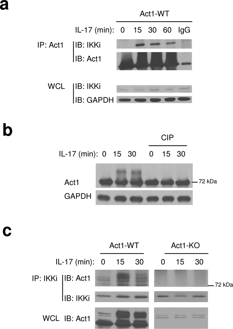
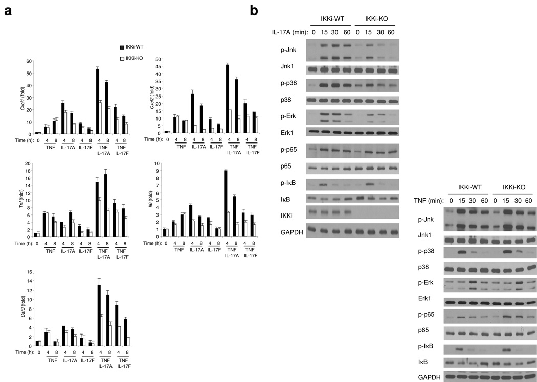
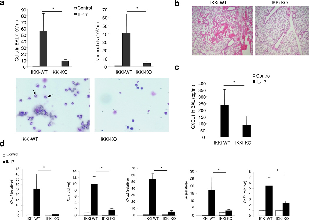
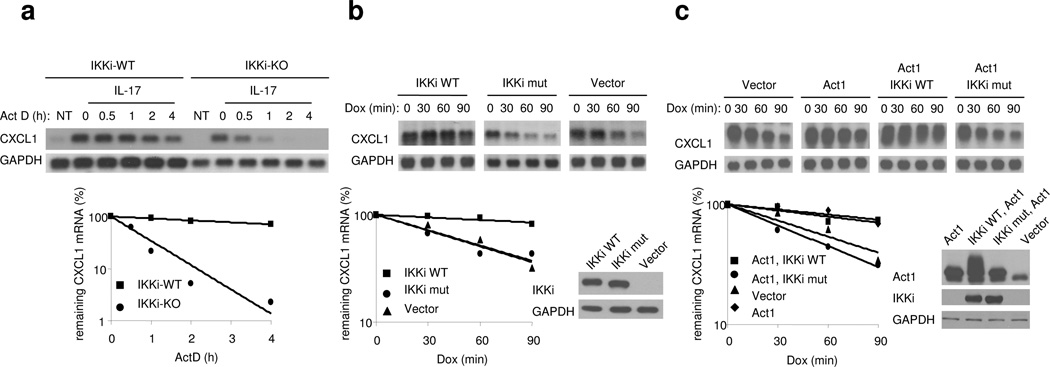
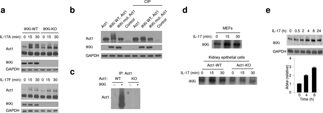
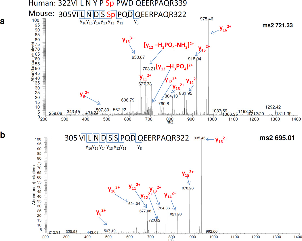
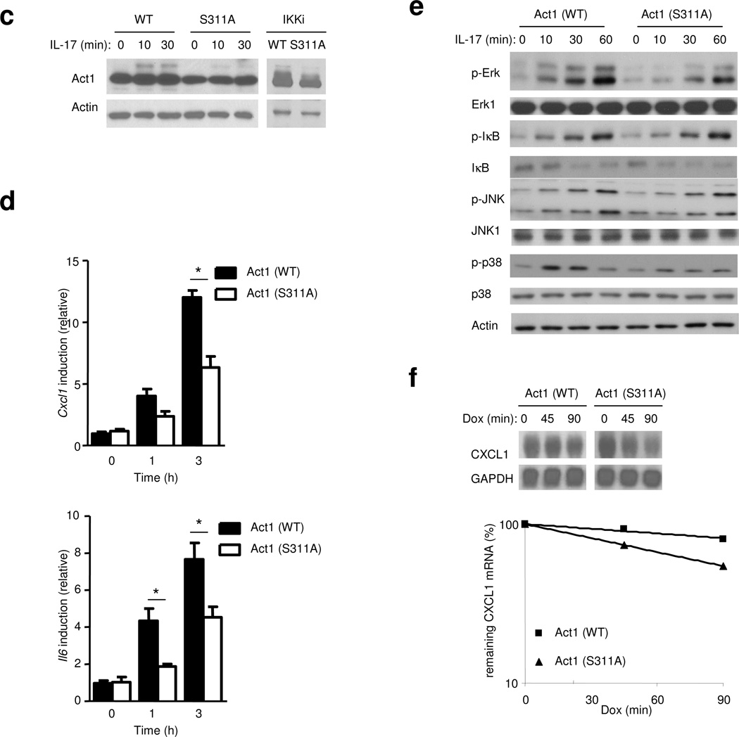
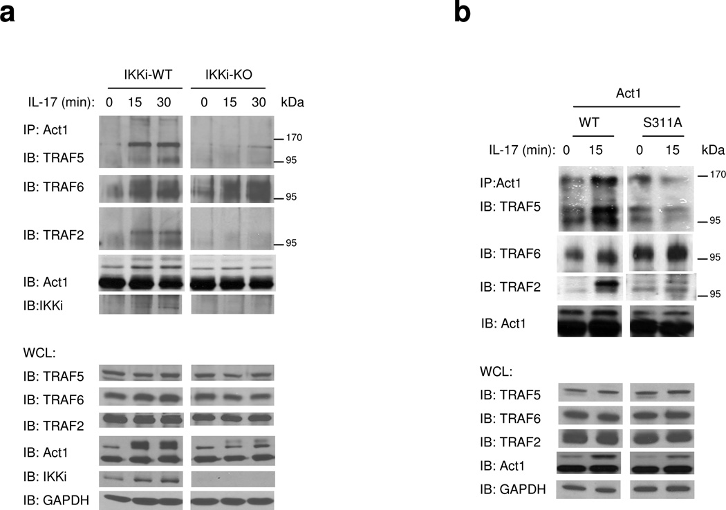
Interleukin 17 (IL-17) is critical in the pathogenesis of inflammatory and autoimmune diseases. Here we report that Act1, the key adaptor for the IL-17 receptor (IL-7R), formed a complex with the inducible kinase IKKi after stimulation with IL-17. Through the use of IKKi-deficient mice, we found that IKKi was required for IL-17-induced expression of genes encoding inflammatory molecules in primary airway epithelial cells, neutrophilia and pulmonary inflammation. IKKi deficiency abolished IL-17-induced formation of the complex of Act1 and the adaptors TRAF2 and TRAF5, activation of mitogen-activated protein kinases (MAPKs) and mRNA stability, whereas the Act1-TRAF6-transcription factor NF-κB axis was retained. IKKi was required for IL-17-induced phosphorylation of Act1 on Ser311, adjacent to a putative TRAF-binding motif. Substitution of the serine at position 311 with alanine impaired the IL-17-mediated Act1-TRAF2-TRAF5 interaction and gene expression. Thus, IKKi is a kinase newly identified as modulating IL-17 signaling through its effect on Act1 phosphorylation and consequent function.


Figures

Figure 1
Figure 1. IKKi forms a complex with Act1 upon IL-17 stimulation
A. Cell lysates from Act1-deficient MEFs infected with retroviral WT Act1 (Act1-WT) untreated or treated with IL-17 (50 ng/ml) for 0, 15, 30 and 60 min were immunoprecipitated with anti-Act1 or IgG, followed by immunoblot analysis with anti-IKKi, anti-Act1 and anti-GAPDH. WCL: whole cell lysates. B. Lysates from Act1 WT reconstituted MEFs treated with IL-17 (50 ng/ml) for 0, 15 and 30 min were untreated or treated with phosphatase (CIP, 1 h, 37°C)], followed by immunoblot analysis with anti-Act1 and anti-GAPDH. C. Cell lysates from wild-type and Act1-deficient MEFs untreated or treated with IL-17 (50 ng/ml) for 0, 15 and 30 min were immunoprecipitated with anti-IKKi, followed by immunoblot analysis with anti-Act1 and anti-IKKi. WCL: whole cell lysates. The data shown in this figure are representation of three independent experiments.
Figure 2
Figure 2. IKKi is required for IL-17-mediated pro-inflammatory gene expression
A. Real-time PCR analysis of CXCL1, TNF, CXCL2, IL-6 and CSF3 in wild-type and IKKi-deficient airway epithelial cells untreated or stimulated with TNF (10ng/ml), IL-17A (50 ng/ml), IL-17F (50 ng/ml), TNF+IL-17A or TNF+IL-17F at the basal surface of the epithelial cells for 0, 4 and 8 hours. Expression of mRNA is presented as fold of induction. B. Immunoblot analysis of p-Jnk, Jnk1, p-p38, p38, p-Erk, Erk1, p-p65, p65, p-IκB, IκB, IKKi and GAPDH of lysates from wild-type and IKKi-deficient airway epithelial cells untreated or treated with IL-17A (50 ng/ml) or with TNF (10ng/ml) for indicated times. The data shown in this figure are representation of three independent experiments.
Figure 3
Figure 3. IKKi is required for IL-17-mediated pulmonary inflammation
A. Total BAL and differential cell count were analyzed in samples from control or IL-17 challenged (1 µg through intranasal injection for 24 h) wild-type and IKKi-deficient mice (n=6) P<0.05. Cytospins prepared from the BAL of control or IL-17 challenged wild-type and IKKi-deficient mice were stained with Hema3, and differential cell counting was performed using standard morphological criteria. Magnification x400. B. The lung sections from control or IL-17 challenged wild-type and IKKi-deficient mice (1 µg through intranasal injection for 24 h) were stained with H&E. Magnification x100. C. ELISA of CXCL1 chemokine in BAL fluid from control or IL-17 challenged wild-type and IKKi-deficient mice. D. Real-time PCR analysis of CXCL1, TNF, CXCL2, IL-6 and CSF3 in the lung tissues from control or IL-17 treated wild-type and IKKi-deficient mice. Expression of mRNA is presented as arbitrary units (mean ± s.d.) relative to the expression of mRNA encoding β-Actin. P<0.05. The data shown in this figure are representation of two independent experiments.
Figure 4
Figure 4. The kinase activity of IKKi is required for chemokine mRNA stabilization
A. Wild-type and IKKi-deficient MEFs were pretreated with TNF (10 ng/ml) for 1 h, followed by the treatment with IL-17 and ActD (5 µg/ml) for the indicated times. Total RNAs were prepared from these cells, followed by analysis of CXCL1 and GAPDH mRNA abundance by RNA hybridization blot analysis. B. HeLa tet-off cells were transfected with 1 µg of pTRE2 CXCL1Δ4 and 1 µg of pcDNA3, IKKi WT or K38A IKKi mutant. The transfected cells were treated with Dox (1 µg/ml) and incubated for the indicated times, followed by analysis of CXCL1 and GAPDH mRNA levels by RNA hybridization blot analysis. C. HeLa tet-off cells were transfected with 1 µg of pTRE2 CXCL1Δ4 and 1 µg of pcDNA3, Act1+ IKKi WT, or Act1+K38A IKKi mutant. The transfected cells were treated with Dox (1 µg/ml) and incubated for the indicated times, followed by analysis of CXCL1 and GAPDH mRNA levels by RNA hybridization blot analysis. The data shown in this figure are representation of three independent experiments.
Figure 5
Figure 5. The kinase activity of IKKi is required for IL-17-induced Act1 phosphorylation
A. Immunoblot analysis of Act1, IKKi and GAPDH in lysates from wild type and IKKi-deficient airway epithelial cells untreated or treated with IL-17A (50 ng/ml) or IL-17F (50ng/ml) for indicated times. B. Lysates from 293HEK cells transfected with Act1+pcDNA3, Act1+ IKKi WT, Act1+K38A IKKi mutant or pcDNA3 were untreated or treated with phosphatase (CIP, 1 h at 37°C), followed by immunoblot analysis with antibodies against Act1, IKKi and GAPDH. C. Lysates from wild-type or Act1-deficient MEFs untreated or treated with IL-17 (50 ng/ml, 20 min) were immunoprecipitated with anti-Act1 followed by in vitro kinase assay with or without recombinant IKKi-GST. D. Lysates from wild-type MEFs or wild-type and Act1-deficient kidney epithelial cells untreated or treated with IL-17 (50 ng/ml, 0, 15 and 30 min) were immunoprecipitated with anti-IKKi followed by in vitro kinase assay. E. Immunoblot and real-time PCR analysis of IKKi and GAPDH in lysates from wild type and IKKi-deficient airway epithelial cells untreated or treated with IL-17A (50 ng/ml) for indicated times. The data shown in this figure are representation of three independent experiments.
Figure 6
Figure 6. Identification of S311 phosphorylation site of Act1 by MS
A. MS/MS spectrum of the Act1 phosphopeptide (305VILNDSSpPQDEERPAQR322) precursor ions at m/z of 721.33 Da. B. MS/MS spectrum of unmodified peptide of the same sequence (305VILNDSSPQDEERPAQR322) precursor ions at m/z of 695.01 Da. C. Act1-deficient MEFs infected with either retroviral WT-mAct1 or Act1 mutant S11A were untreated or treated with IL-17 (50 ng/ml) for 0, 15 or 30 min, followed by immunoblot analysis with anti-Act1 and anti-Actin. D. Act1-deficient MEFs infected with either retroviral WT-mAct1 or Act1 mutant S11A, followed by treatments with IL-17A (50 ng/ml) for 1 h and 3 h. CXCL1 and IL-6 mRNA was measured by real-time PCR. E. Act1-MEFs infected either WT-mAct1 or Act1 mutant S11A were untreated or treated with IL-17 (50 ng/ml) for 0, 15, 30 or 60 min, followed by immunoblot analysis with antibodies against p-Erk, Erk1, p-IκB, IκB, p-Jnk, Jnk, p-p38, p38 and Actin. F. HeLa tet-off cells were transfected with 1 µg of pTRE2 CXCL1Δ4 and 1 µg of Act1 or Act1 S311A mutant. The transfected cells were treated with dox (1 µg/ml) and incubated for the indicated times, followed by isolation of total RNA and RNA hybridization blot analysis for the determination of CXCL1 and GAPDH mRNA levels. The data shown in this figure are representation of three independent experiments.
Figure 7
Figure 7. The impact of phosphorylation of Act1-S311 on Act1’s interaction with TRAFs
A. Cell lysates from wild type and IKKi-deficient kidney epithelial cells untreated or treated with IL-17 (50 ng/ml) for 0, 15 and 30 min were immunoprecipitated with anti-Act1, followed by immunoblot analysis with antibodies against TRAF2, TRAF5, TRAF6, IKKi and Act1. WCL (whole cell lysates). The data shown is a representation of three independent experiments. B. Act1-deficient MEFs infected with either retroviral WT-mAct1 or Act1 mutant S11A were untreated or treated with IL-17A (50 ng/ml) for 0 or 15 min. Cell lysates were then immunoprecipitated with anti-Act1, followed by immunoblot analysis with antibodies against TRAF2, TRAF5, TRAF6 and Act1. WCL (whole cell lysates). The data shown is a representation of three independent experiments. C. Computationally modeled interaction of the Act1 TRAF-binding motif (human Act1327–334) to the structures of TRAF domains of TRAF6 (yellow) and TRAF2 (pink) and a homology model of the TRAF domain of TRAF5 (orange). The Act1 target Serine/phosphoserine is marked with a dashed circle in each panel. D. –F. Details of representative models of the Act1 TRAF-binding motifs docked to TRAF domains of (D) TRAF6, (E) TRAF5 and (F) TRAF2 respectively. Act1-interacting sidechains of the TRAF domains are shown and labeled in standard text. Residues of Act1 are labeled in bold, while the key Act1 Serine/phosphoserine is labeled in red bold. Putative hydrogen bonds or salt bridges between Act1 and TRAF domain sidechains are shown as dashed lines. Hydrogen bonds between Act1 backbone atoms and TRAF domain backbone atoms are also shown as dashed lines but, for clarity, the corresponding TRAF domain backbone atoms are not shown explicitly.

Comment in

References

    1. Bettelli E, et al. Reciprocal developmental pathways for the generation of pathogenic effector TH17 and regulatory T cells. Nature. 2006;441:235–238. - PubMed
    1. Cho JS, et al. IL-17 is essential for host defense against cutaneous Staphylococcus aureus infection in mice. J. Clin. Invest. 2010;120:1762–1773. - PMC - PubMed
    1. Conti HR, et al. Th17 cells and IL-17 receptor signaling are essential for mucosal host defense against oral candidiasis. J. Exp. Med. 2009;206:299–311. - PMC - PubMed
    1. Harrington LE, et al. Interleukin 17-producing CD4+ effector T cells develop via a lineage distinct from the T helper type 1 and 2 lineages. Nat. Immunol. 2005;6:1123–1132. - PubMed
    1. Iwakura Y, Ishigame H. The IL-23/IL-17 axis in inflammation. J. Clin. Invest. 2006;116:1218–1222. - PMC - PubMed

Publication types

MeSH terms