. 2011 Aug 7;12(9):853-60.
doi: 10.1038/ni.2081.

Treatment with IL-17 prolongs the half-life of chemokine CXCL1 mRNA via the adaptor TRAF5 and the splicing-regulatory factor SF2 (ASF)

Affiliations

Treatment with IL-17 prolongs the half-life of chemokine CXCL1 mRNA via the adaptor TRAF5 and the splicing-regulatory factor SF2 (ASF)

Dongxu Sun et al. Nat Immunol. .

Abstract

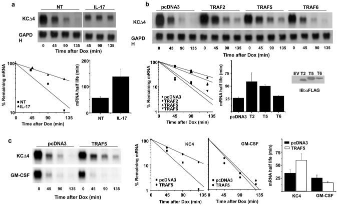
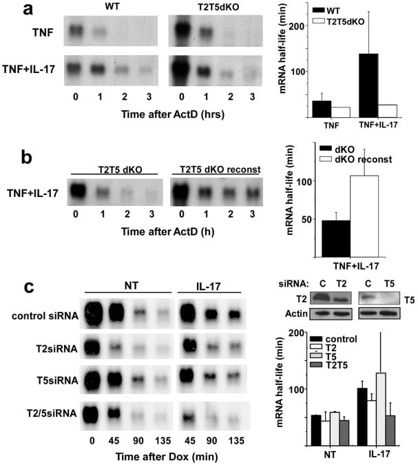
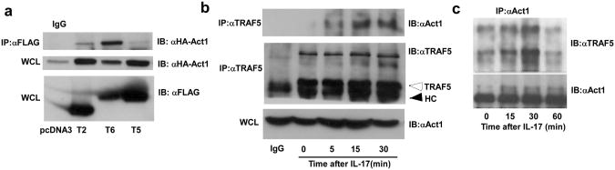
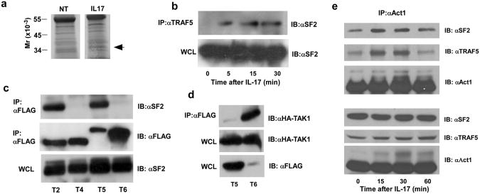
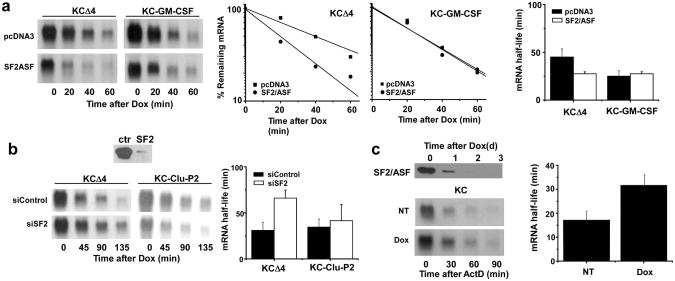
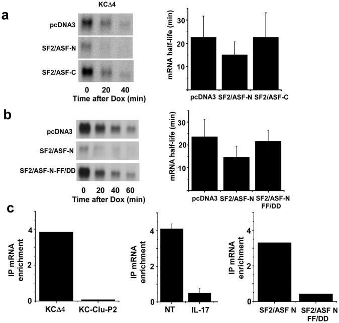
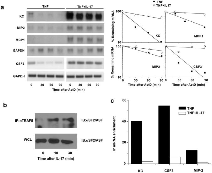
Interleukin 17 (IL-17) promotes the expression of chemokines and cytokines via the induction of gene transcription and post-transcriptional stabilization of mRNA. We show here that IL-17 enhanced the stability of chemokine CXCL1 mRNA and other mRNAs through a pathway that involved the adaptor Act1, the adaptors TRAF2 or TRAF5 and the splicing factor SF2 (also known as alternative splicing factor (ASF)). TRAF2 and TRAF5 were necessary for IL-17 to signal the stabilization of CXCL1 mRNA. Furthermore, IL-17 promoted the formation of complexes of TRAF5-TRAF2, Act1 and SF2 (ASF). Overexpression of SF2 (ASF) shortened the half-life of CXCL1 mRNA, whereas depletion of SF2 (ASF) prolonged it. SF2 (ASF) bound chemokine mRNA in unstimulated cells, whereas the SF2 (ASF)-mRNA interaction was much lower after stimulation with IL-17. Our findings define an IL-17-induced signaling pathway that links to the stabilization of selected mRNA species through Act1, TRAF2-TRAF5 and the RNA-binding protein SF2 (ASF).


Figures

Figure 1
Figure 1. TRAF2 or TRAF5 expression selectively prolongs CXCL1 mRNA half life
a. HeLa Tet-off cells were transfected with the reporter plasmid KCΔ4 and treated with dox alone or in the presence of IL-17 (25 ng/ml) for the indicated times prior to quantification of residual CXCL1 and GAPDH mRNA by RNA hybridization. The autoradiograph was scanned and quantified using ImageJ software and the abundance of CXCL1 mRNA was normalized to GAPDH mRNA in each sample. The results of 3 independent experiments were used to determine the half-lives under resting and stimulated conditions and are presented as the mean +/- 1 S.D. b. HeLa tet-off cells were co-transfected with KCΔ4 and vector alone (pcDNA3) or plasmids encoding TRAF2, TRAF5, or TRAF6. Cultures were treated with dox (1 μg/ml) for the indicated times prior to quantification of individual TRAFs by immunoblot and CXCL1 mRNA as described in a. Half-lives were calculated from 3 independent experiments. c. HeLa tet-off cells were co-transfected with reporter plasmids KCΔ4 or KC-GM-CSF along with pcDNA3 or the TRAF5 expression plasmid. Dox was added and the remaining CXCL1 and GAPDH (not shown) mRNA was quantified and half-lives determined from 3 independent experiments as described in a.
Figure 2
Figure 2. TRAF2 and TRAF5 are required for IL-17-induced CXCL1 mRNA stabilization
a. Wild type and TRAF2-/-,TRAF5-/- MEFs were treated with TNF (10 ng/ml) alone or in the presence of IL-17 for 2 h prior to the addition of ActD (5 μg/ml). Remaining CXCL1 and GAPDH (not shown) mRNA was determined by RNA hybridization and the half-lives calculated as described in the legend to Figure 1a from 4 independent experiments. b. TRAF2/TRAF5 deficient MEFs and TRAF-reconstituted MEFs were treated with TNF+IL-17 (10 ng/ml each) for 2 hrs prior to addition of ActD (5 μg/ml) and remaining CXCL1 and GAPDH (not shown) mRNA was quantified and half-lives calculated as in figure 1 from 3 independent experiments. c. HeLa tet-off cells were co-transfected with control siRNA or siRNA specific for TRAF2 or TRAF5 along with the KCΔ4 reporter. Separate cultures were used to determine residual TRAF expression by immunoblot. The cells were treated with dox alone or along with IL-17 and CXCL1 and GAPDH (not shown) mRNA was quantified and half-lives calculated as described in a. Similar results were obtained in 2 independent experiments.
Figure 3
Figure 3. IL-17 promotes interaction of Act1 and TRAF5
a. HeLa tet-off cells were co-transfected with FLAG-TRAF2, FLAG-TRAF5, or FLAG-TRAF6 along with HA-Act1 used for immunoprecipitation with FLAG antibody. The IPs were analyzed by SDS-PAGE and immunoblot with anti-HA. 40 μg protein from whole cell lysates (WCL) were analyzed by immunoblot using anti-FLAG to illustrate TRAF expression. Results are representative of 3 independent experiments. b. HeLa tet-off cells were treated with IL-17 for the indicated times and used for immunoprecipitation with TRAF5 antibody. The IPs were analyzed by SDS-PAGE and immunoblot with anti-Act1 or anti-TRAF5. The control IgG lane used WCL from cells treated with IL-17 for 5 minutes. 40 μg of each WCL were subjected to immunoblot for Act1. The unfilled arrow indicates the position of unmodified TRAF5 and the solid arrow indicates the position of antibody heavy chain (HC). Results are representative of 2 independent experiments. c. Wild type MEFs were treated with IL-17 for the indicated times and used for immunoprecipitation with anti-Act1. The IPs were analyzed by SDS-PAGE and immunoblot with anti-TRAF5 or anti-Act1. The results are representative of 3 independent experiments.
Figure 4
Figure 4. IL-17 induces a complex with TRAF2 or TRAF5 and SF2/ASF
a. MEFs from TRAF2/TRAF5-deficient mice that were reconstituted with FLAG-TRAF2 and FLAG-TRAF5 were untreated or stimulated with IL-17 (10 ng/ml) for 1 hr. Cell lysates were immunoprecipitated with anti-FLAG and the IPs were subjected to SDS-PAGE followed by silver staining. The band identified by the filled arrow was subjected to in-gel trypsin digestion and LC-MS and the data analyzed using MASCOT software to search the NCBI non-redundant data base. b. HeLa tet-off cells were untreated or treated with IL-17 (25 ng/ml) for the indicated times prior to immunoprecipitation with anti-TRAF5. The IPs or 40 μg of WCL protein were analyzed by SDS-PAGE and immunoblot using anti-SF2/ASF. Results are representative of 2 independent experiments. c. HeLa tet-off cells were transfected to express FLAG-tagged versions of TRAF2, TRAF4, TRAF5 or TRAF6 and used for immunoprecipitation with anti-FLAG. The IPs or 40 μg WCL protein were analyzed by SDS-PAGE and immunoblot using anti-SF2/ASF or anti-FLAG to detect TRAFs. d. HeLa tet-off cells were co-transfected to express FLAG-TRAF5 or FLAG-TRAF6 along with HA-TAK1 and used for immunoprecipitation with anti-FLAG. IPs or 40 μg WCL protein were analyzed by SDS-PAGE and immunoblot using anti-HA for TAK1 and anti-FLAG for TRAFs. Results are representative of 2 independent experiments. e. Extracts from HeLa tet-off cells treated with IL-17 (50 ng/ml) for the indicated times immunoprecipitated with anti-Act1. The IPs and wcl extracts were analyzed by SDS-PAGE and immunoblot using antibodies to SF2/ASF, TRAF5, and Act1. Results are representative of 3 independent experiments.
Figure 5
Figure 5. SF2/ASF promotes enhanced decay of CXCL1 mRNA
a. Hela tet-off cells were co-transfected with KCΔ4 or KC-GM-CSF and either vector alone (pcDNA3) or full length SF2/ASF. Dox (1 μg/ml) was added and residual CXCL1 and GAPDH (not shown) mRNA was quantified by RNA hybridization. Half-lives were calculated as described in the legend to figure 1 from 3 independent experiments. b. Hela tet-off cells were co-transfected with reporter plasmids KCΔ4 or KC-Clu-P2 along with control or SF2/ASF-specific siRNA. After 48 hrs, cells were treated with Dox (1 μg/ml) and CXCL1 and GAPDH (not shown) mRNA was quantified by RNA hybridization and half lives were calculated as in the legend to figure 1 from 2 independent experiments. 40 μg of WCL was used to measure SF2/ASF expression by immunoblot analysis. c. SF2/ASF knockout MEFs reconstituted with HA-tagged SF2/ASF under tet-off control were untreated or treated with Dox (1 μg/ml) for 2 days prior to stimulation with TNF (10 ng/ml) for 2 hrs followed by ActD (5 μg/ml) for the indicated times. CXCL1 and GAPDH (not shown) mRNA was measured by RNA hybridization and half-lives were calculated as in the legend to figure 1 from 2 independent experiments. HA-SF2/ASF was quantified in untreated and Dox-treated cells by immunblot using 40 μg WCL protein.
Figure 6
Figure 6. SF2/ASF binds CXCL1 mRNA
a. HeLa tet-off cells were co-transfected with KCΔ4 and expression plasmids encoding empty vector (pcDNA3), C-terminal deletion (SF2/ASF-N), or N-terminal deletion (SF2/ASF-C) versions of SF2/ASF. Dox (1 μg/ml) was added and remaining CXCL1 and GAPDH (not shown) mRNA was quantified and half-lives were calculated as described in the legend to figure 1 using results from 3 independent experiments. b. HeLa tet-off cells were co-transfected with KCΔ4 reporter plasmid and vector alone (pcDNA3) or pcDNA3 encoding either the wild-type (SF2/ASF-N) or mutant (SF2/ASF-N-FF-DD) C terminal deletion variant of SF2/ASF. Dox (1 μg/ml) was added and remaining CXCL1 or GAPDH (not shown) mRNA was quantified and half-lives were calculated as in the legend to figure 1 using results from 2 independent experiments. c. HeLa tet-off cells were transfected with KCΔ4 or KC-Clu-P2 plasmids with or without either the wild-type or RNA binding mutant (FF-DD) version of SF2/ASF C-terminal deletion as indicated. In some experiments the cells were either untreated or treated with IL-17 for 30 min. Extracts were prepared and used for RNA immunoprecipitation with anti-SF2/ASF or anti-HA (for cells expressing the transgenic versions of SF2/ASF). RNA was prepared from IPs and used to quantify CXCL1 and GAPDH mRNA by real time RT-PCR. The data are presented as mRNA enrichment comparing ΔΔCt value for non-specific IgG with that for anti-SF2/ASF. Data are presented as the mean fold enrichment +/- ½ the range of two independent experiments.
Figure 7
Figure 7. IL-17 promotes TRAF5 and SF2/ASF function in primary cells
a. Primary kidney epithelial cells were treated with TNF (10 ng/ml) alone or in combination with IL-17 (25 ng/ml) for 2 hrs. ActD (5 μg/ml) was added to each dish for the indicated times and CXCL1, MIP-2, MCP-1, and GAPDH mRNA was quantified by RNA hybridization. CSF3 and GAPDH mRNA was quantified in the same total RNA samples using RT-PCR (27 cycles) for greater sensitivity. Results are representative of 2 independent experiments. b. Primary kidney epithelial cells were treated with nothing or with IL-17 (25 ng/ml) for the indicated times and used for immunoprecipitation with anti-TRAF5. The IPs or 40 μg of WCL protein were analyzed by SDS-PAGE immunoblot using anti-SF2/ASF. Results are representative of 2 separate experiments. c. Primary kidney epithelial cells were treated with TNF for 2 hrs. Separate cultures were then left untreated or treated with IL-17 (25 ng/ml) for 30 minutes. Extracts were used for RNA immunoprecipitation with anti-SF2/ASF. RNA was recovered from IPs and used to quantify CXCL1, CSF3, MIP-2 and GAPDH mRNAs by real time RT-PCR. The data are presented as mRNA enrichment comparing ΔΔCt value for non-specific IgG with that for anti-SF2/ASF. Results are representative of 3 independent experiments.

Comment in

References

    1. Ouyang W, Kolls JK, Zheng Y. The biological functions of T helper 17 cell effector cytokines in inflammation. Immunity. 2008;28:454–467. - PMC - PubMed
    1. Korn T, Bettelli E, Oukka M, Kuchroo VK. IL-17 and Th17 Cells. Annu Rev Immunol. 2009;27:485–517. - PubMed
    1. Kang Z, et al. Astrocyte-restricted ablation of interleukin-17-induced Act1-mediated signaling ameliorates autoimmune encephalomyelitis. Immunity. 2010;32:414–425. - PMC - PubMed
    1. Endlich B, Armstrong D, Brodsky J, Novotny M, Hamilton TA. Distinct temporal patterns of macrophage-inflammatory protein-2 and KC chemokine gene expression in surgical injury. J Immunol. 2002;168:3586–3594. - PubMed
    1. Kobayashi Y. Neutrophil infiltration and chemokines. Crit Rev Immunol. 2006;26:307–16. - PubMed

Publication types

MeSH terms