

Natural products reveal cancer cell dependence on oxysterol-binding proteins
- PMID: 21822274
- PMCID: PMC3158287
- DOI: 10.1038/nchembio.625
Natural products reveal cancer cell dependence on oxysterol-binding proteins
Abstract
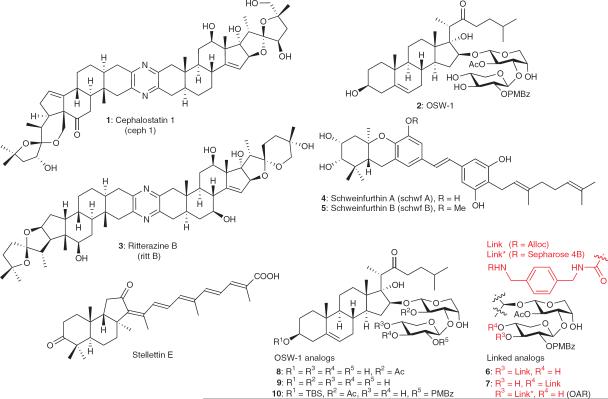
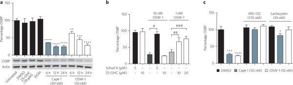
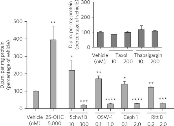
Cephalostatin 1, OSW-1, ritterazine B and schweinfurthin A are natural products that potently, and in some cases selectively, inhibit the growth of cultured human cancer cell lines. The cellular targets of these small molecules have yet to be identified. We have discovered that these molecules target oxysterol binding protein (OSBP) and its closest paralog, OSBP-related protein 4L (ORP4L)--proteins not known to be involved in cancer cell survival. OSBP and the ORPs constitute an evolutionarily conserved protein superfamily, members of which have been implicated in signal transduction, lipid transport and lipid metabolism. The functions of OSBP and the ORPs, however, remain largely enigmatic. Based on our findings, we have named the aforementioned natural products ORPphilins. Here we used ORPphilins to reveal new cellular activities of OSBP. The ORPphilins are powerful probes of OSBP and ORP4L that will be useful in uncovering their cellular functions and their roles in human diseases.
Figures

Similar articles
-
Molecular and cellular dissection of the oxysterol-binding protein cycle through a fluorescent inhibitor.J Biol Chem. 2020 Mar 27;295(13):4277-4288. doi: 10.1074/jbc.RA119.012012. Epub 2020 Feb 19. J Biol Chem. 2020. PMID: 32075908 Free PMC article.
-
Oxysterol-binding proteins: sterol and phosphoinositide sensors coordinating transport, signaling and metabolism.Prog Lipid Res. 2013 Oct;52(4):529-38. doi: 10.1016/j.plipres.2013.06.004. Epub 2013 Jul 2. Prog Lipid Res. 2013. PMID: 23830809 Review.
-
Broad-range inhibition of enterovirus replication by OSW-1, a natural compound targeting OSBP.Antiviral Res. 2015 May;117:110-4. doi: 10.1016/j.antiviral.2015.02.013. Epub 2015 Mar 6. Antiviral Res. 2015. PMID: 25752737
-
The role of oxysterol-binding protein and its related proteins in cancer.Semin Cell Dev Biol. 2018 Sep;81:149-153. doi: 10.1016/j.semcdb.2017.07.017. Epub 2017 Jul 18. Semin Cell Dev Biol. 2018. PMID: 28733164 Review.
-
Oxysterol-binding proteins: functions in cell regulation beyond lipid metabolism.Biochem Pharmacol. 2013 Jul 1;86(1):89-95. doi: 10.1016/j.bcp.2013.02.016. Epub 2013 Feb 18. Biochem Pharmacol. 2013. PMID: 23428468 Review.
Cited by
-
Ligand-dependent localization and function of ORP-VAP complexes at membrane contact sites.Cell Mol Life Sci. 2015 May;72(10):1967-87. doi: 10.1007/s00018-014-1786-x. Epub 2014 Nov 25. Cell Mol Life Sci. 2015. PMID: 25420878 Free PMC article.
-
Golgi-Targeting Anticancer Natural Products.Cancers (Basel). 2023 Mar 31;15(7):2086. doi: 10.3390/cancers15072086. Cancers (Basel). 2023. PMID: 37046746 Free PMC article. Review.
-
Functions of Oxysterol-Binding Proteins at Membrane Contact Sites and Their Control by Phosphoinositide Metabolism.Front Cell Dev Biol. 2021 Jun 24;9:664788. doi: 10.3389/fcell.2021.664788. eCollection 2021. Front Cell Dev Biol. 2021. PMID: 34249917 Free PMC article. Review.
-
Lipid transfer proteins rectify inter-organelle flux and accurately deliver lipids at membrane contact sites.J Lipid Res. 2018 Aug;59(8):1341-1366. doi: 10.1194/jlr.R085324. Epub 2018 Jun 8. J Lipid Res. 2018. PMID: 29884707 Free PMC article. Review.
-
Modulation of the Host Lipid Landscape to Promote RNA Virus Replication: The Picornavirus Encephalomyocarditis Virus Converges on the Pathway Used by Hepatitis C Virus.PLoS Pathog. 2015 Sep 25;11(9):e1005185. doi: 10.1371/journal.ppat.1005185. eCollection 2015 Sep. PLoS Pathog. 2015. PMID: 26406250 Free PMC article.
References
-
- Pettit GR, et al. Antineoplastic agents. 147. Isolation and structure of the powerful cell growth inhibitor cephalostatin 1. J. Am. Chem. Soc. 1988;110:2006–2007.
-
- Kubo S, et al. Acylated cholestane glycosides from the bulbs of Ornithogalum saundersiae. Phytochemistry. 1992;31:3969–3973.
-
- Komiya T, et al. Ritterazine B, a new cytotoxic natural compound, induces apoptosis in cancer cells. Cancer Chemother. Pharmacol. 2003;51:202–208. - PubMed
-
- Beutler JA, Shoemaker RH, Johnson T, Boyd MR. Cytotoxic geranyl stilbenes from Macaranga schweinfurthii. J. Nat. Prod. 1998;61:1509–1512. - PubMed
-
- Tasdemir D, et al. Bioactive isomalabaricane triterpenes from the marine sponge Rhabdastrella globostellata. J. Nat. Prod. 2002;65:210–214. - PubMed
Publication types
MeSH terms
Substances
Associated data
Grants and funding
LinkOut - more resources
Full Text Sources
Other Literature Sources
