

Review
doi: 10.1038/nrmicro2622.
Function, structure and mechanism of bacterial photosensory LOV proteins
Affiliations
- PMID: 21822294
- PMCID: PMC3286519
- DOI: 10.1038/nrmicro2622
Item in Clipboard
Review
Function, structure and mechanism of bacterial photosensory LOV proteins
Nat Rev Microbiol.
.
Abstract
LOV (light, oxygen or voltage) domains are protein photosensors that are conserved in bacteria, archaea, plants and fungi, and detect blue light via a flavin cofactor. LOV domains are present in both chemotrophic and phototrophic bacterial species, in which they are found amino-terminally of signalling and regulatory domains such as sensor histidine kinases, diguanylate cyclases-phosphodiesterases, DNA-binding domains and regulators of RNA polymerase σ-factors. In this Review, we describe the current state of knowledge about the function of bacterial LOV proteins, the structural basis of LOV domain-mediated signal transduction, and the use of LOV domains as genetically encoded photoswitches in synthetic biology.
Figures
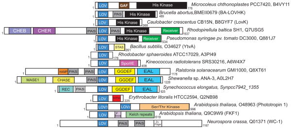
Domain architecture of select LOV proteins; this cartoon presents just a few examples of hundreds of LOV domain-containing proteins. Accession numbers and formal protein names (if available) are shown. The length of LOV proteins is specified.
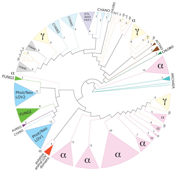
LOV domain sequences are conserved across multiple kingdoms. Tree adapted from Krauss and colleagues . LOV sequences of different families are color coded: the archaea in turquoise, alphaproteobacteria (α) in pink, betaproteobacteria (β) and gammaproteobacteria (γ) in yellow, actinobacteria (actino) are in brown, chloroflexi (chloro) in dark green, firmicutes (firmi) in grey, cyanobacteria (cyano) in light blue, fungal sequences from the white-collar 1 (WC-1) protein are in green (fungi), plant phototropin and neochrome (Phot/Neo) LOV domains (LOV1 and LOV2) are in blue, algal aureochrome LOV sequences (aureo) in dark purple, the ZTL/ADO/FKF1-LOV family of plant regulators are in purple, and putative animal LOV domains are in red. Branches have been collapsed; triangle size is proportional to the number of collapsed branches. Numbers of collapsed branches are indicated at the base of each triangle.
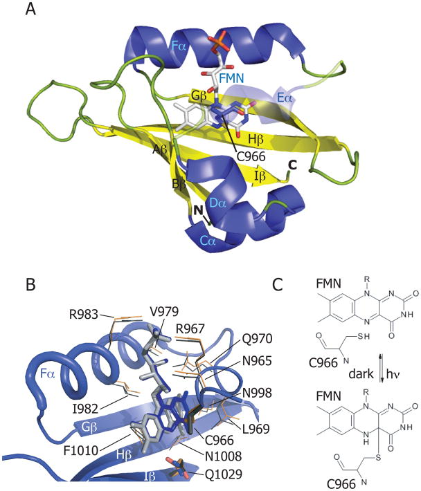
(A) Adiantum capillus-veneris phy3(neochrome)-LOV2 domain in the illuminated state (PDB code: 1JNU). α-helices are colored in blue; β-strands in yellow; loops in green. The FMN cofactor is colored in light gray. Helix Eα is shown as transparent to reveal the cysteinyl-FMN adduct. (B) Flavin-binding pocket of phy3-LOV2 domain. Residues that interact with the FMN cofactor are shown for the illuminated state (PDB code: 1JNU) (in orange), and for the dark state (PDB code: 1G28) (in gray). The glutamine (Q1029) of strand Hβ that rotates 180° on light activation is labeled as is the cysteine that forms the cysteinyl-FMN adduct (C966). (C) Schematic of cysteinyl-C(4a) covalent adduct formation in response to light absorption by the LOV domain.
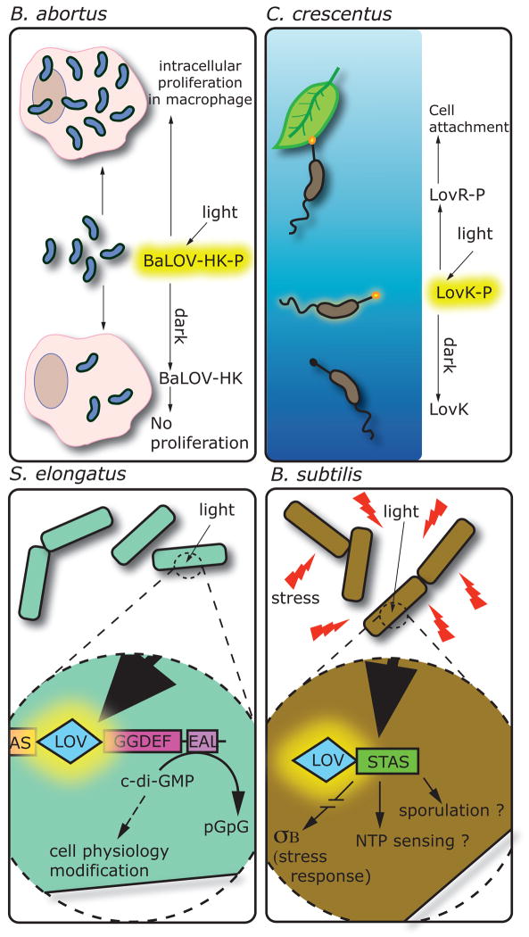
Biochemical activities and cellular responses affected by LOV photoreceptor proteins in four bacterial species. Brucella abortus: on light activation BaLOV-HK is phosphorylated and positively regulates intracellular proliferation in a macrophage infection model. Caulobacter crescentus: light activation of the LovK protein induces autophophorylation. LovK, together with LovR, regulates cell adhesion through an unknown mechanism. Synechoccocus elongatus: regulation of the phosphodiesterase activity of the EAL domain is controlled by the LOV domain. Thus, light exposure controls the di-c-GMP level of the cell, an important second messenger. Bacillus subtilis: during stress conditions, illumination of the LOV domain of the YtvA protein is an important signal for σB activation and stress response. The associated STAS domain, maybe involved in energy level sensing, could also be regulated by the LOV domain as well as sporulation phenomenon.
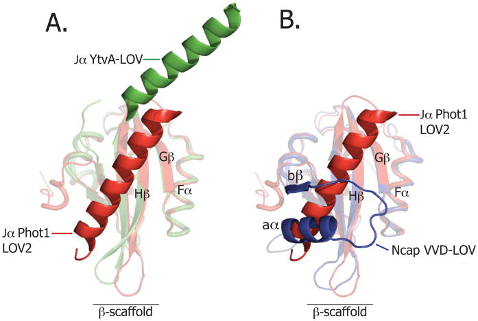
(A) Structural alignment of the dark-state structure of the YtvA LOV domain of B. subtilis (in green; PDB code: 2PR5) and dark-state phototropin 1 LOV domain of A. sativa (in red; PDB code: 2V0U). (B) Structural alignment of the dark state VVD LOV domain of N. crassa (in blue, PDB code: 2PD7) and the dark Phototropin 1 LOV domain of A. sativa (in red, PDB code: 2V0U).
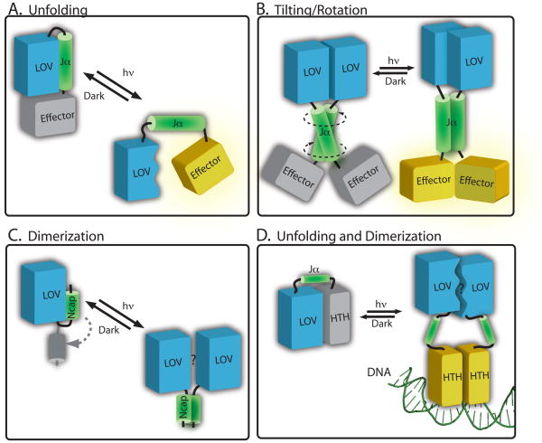
Structural models of signalling in LOV proteins (A) In phototropin-type signaling, cysteinyl-flavin adduct formation induces conformational change in the LOV2 domain (blue) that results in disruption of the interaction with the Jα helix (green). This leads to effector domain activation (yellow). Data from YtvA and bacterial LOV histidine kinases evidence models in which illumination of a LOV domain (blue) can induce conformational changes in an extended Jα helix (green), causing (B) tilting or rotational motion to activate the effector (in yellow). (C) In VVD, light activation leads to rearrangement of the N-terminal cap (in green) and a subsequent change in protein dimerization. (D) In the E. litoralis LOV-HTH protein, EL222, it has been proposed that light activation disrupts the interaction surface between the LOV domain and the HTH domain. This light-driven structural change leads to dimerization of the protein on DNA.
References
-
- Lai EC. RNA sensors and riboswitches: self-regulating messages. Curr Biol. 2003;13:R285–291. - PubMed
Publication types
MeSH terms
Substances
Grants and funding
LinkOut - more resources
Full Text Sources
Other Literature Sources
