. 2012 Mar 22;31(12):1592-8.
doi: 10.1038/onc.2011.349. Epub 2011 Aug 8.

A common single-nucleotide polymorphism in cyclooxygenase-2 disrupts microRNA-mediated regulation

Affiliations

A common single-nucleotide polymorphism in cyclooxygenase-2 disrupts microRNA-mediated regulation

A E Moore et al. Oncogene. .

Abstract

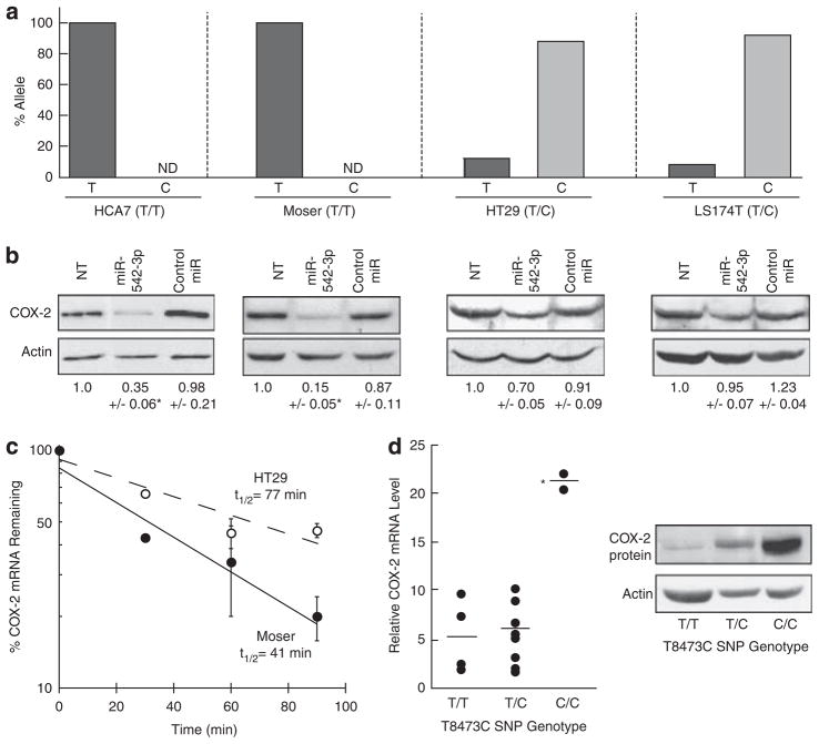
Elevated expression of the prostaglandin synthase cyclooxygenase-2 (COX-2) is commonly observed in many chronic inflammatory diseases and cancer. However, the mechanisms allowing for pathogenic COX-2 overexpression are largely unknown. The gene for COX-2 (PTGS2) carries a common single-nucleotide polymorphism (SNP) at position 8473 (T8473C), in exon 10 that is associated with diseases in which COX-2 overexpression is a contributing factor. We demonstrate that the T8473C SNP resides within a region that targets COX-2 mRNA for degradation through microRNA-mediated regulation. miR-542-3p was identified to bind transcripts derived from the 8473T allele and promote mRNA decay. By contrast, the presence of the variant 8473C allele interfered with miR-542-3p binding, allowing for mRNA stabilization, and this effect was rescued using a mutated miR-542-3p at the respective 8473 site. Colon cancer cells and tissue displayed COX-2 mRNA levels that were dependent on T8473C allele dosage, and allele-specific expression of COX-2 was observed to be a contributing factor promoting COX-2 overexpression. These findings provide a novel molecular explanation underlying disease susceptibility associated with COX-2 T8473C SNP, and identify it as a potential marker for identifying cancer patients best served through selective COX-2 inhibition.


Conflict of interest statement

Conflict of interest

The authors declare no conflict of interest.

Figures

Figure 1
Figure 1
miR-542-3p regulates COX-2 expression. HeLa cells grown in Dulbecco’s modified Eagle’s medium containing 10% fetal bovine serum were treated with 10 ng/ml IL-1β for 24 h to induce COX-2 expression, and then transfected for 48 h with 50 nM hsa-miRNA-542-3p mature miRNA duplex, or random sequence control miRNA #2 (Ambion, Austin, TX, USA) using siQuest (Mirus, Madison, WI, USA). Non-transfected (NT) cells are indicated. (a) Total RNA was isolated using Trizol reagent (Invitrogen, Carlsbad, CA, USA). Northern blotting was performed as described (Dixon et al., 2000), and probed with P32-labeled DNA probes for COX-2 and control actin. (b) cDNA was synthesized from 1 μg total RNA, and Taqman-based quantitative PCR reactions were performed to detect COX-2 mRNA. All values shown are first normalized to control glyceraldehydes 3-phosphate dehydrogenase (GAPDH) and then to the respective NT control. *P=0.0008. (c) COX-2 protein expression was evaluated by immunoblotting as described (Young et al., 2009), using an anti-COX-2 antibody (160112; Cayman, Ann Arbor, MI, USA). Membranes were stripped and re-probed using β-actin (Clone C4; MP Biomedicals, Solon, OH, USA). Detection and quantitation of blots were performed as described (Dixon et al., 2000). Densitometry analysis was used to determine relative fold change in COX-2 intensity relative to the NT control and is listed underneath each representative sample. *P=0.0254. Data shown represents three experiments. (d) COX activity in IL-1β-stimulated HeLa cells transfected with miR-542-3p was assayed by determining PGE2 levels in culture media by ELISA (R&D Systems, Minneapolis, MN, USA). Where indicated, cells were pre-treated with 10 μM NS-398 (Cayman) for 1 h, after which the media was removed and cultures were incubated for 20 min with serum-free media containing 10 μM arachidonic acid (Cayman). Relative PGE2 levels were normalized to total protein levels and are an average of three experiments. *P<0.0001. (e) COX-2 mRNA half-life (t1/2) was determined in HeLa cells stimulated with IL-1β for 24 h and then transfected with miR-542-3p (black circles) or control miR (open circles) for 48 h. Actinomycin D (5 μg/ml) was added to the growth medium and total RNA was prepared at the indicated time points. COX-2 mRNA decay was analyzed by real-time quantitative PCR using Taqman probes (Applied Biosystems) for detection of COX-2, and normalized to control GAPDH as described (Young et al., 2009).
Figure 2
Figure 2
miR-542-3p targets the COX-2 3′UTR. (a, b) HeLa cells treated with IL-1β for 24 h were initially transfected with HA-tagged Ago1 expression plasmid (pHA-Ago1; (Abdelmohsen et al., 2008)), using Lipofectamine Plus (Invitrogen) for 3 h. Media was changed and cells were transfected with either miR-542-3p or control miR, using siQuest for 24 h. Analysis of COX-2 mRNA (a) or miR-542-3p (b) with the RISC complex was performed as described (Sanduja et al., 2009), using a polyclonal anti-HA antibody (Santa Cruz Biotechnology, Santa Cruz, CA, USA) or control IgG pre-coated to protein A/G PLUS agarose (Santa Cruz Biotechnology) to immunoprecipitate HA-tagged Ago1-containing RISC complex. Total RNA was isolated from immunoprecipitates using 1ml Trizol per IP reaction. Taqman-based quantitative PCR reactions were performed to detect COX-2 mRNA or miR-542-3p as described (Sanduja et al., 2009). All values shown are normalized to the respective IgG control miRNA ribonucleoprotein immunoprecipitation reaction, and indicate the averages of three experiments. *P=0.0031; *P=0.0001. (c) HeLa cells treated with IL-1β for 24 h were initially transfected for 24 h with 50 nM siRNA for EIF2C1/Ago-1 or control siRNA (Ambion) using siQuest. Media was changed and cells were transfected with either miR-542-3p or control miR using siQuest for 24 h. Total RNA was isolated and Taqman-based quantitative PCR reactions were performed to detect COX-2 mRNA. *P=0.0007. (d) HeLa cells were transfected sequentially with luciferase (Luc) 3′-UTR reporter constructs (Dixon et al., 2000) containing no 3′-UTR, full-length 8473T-containing COX-2 3′-UTR (Luc+3′-UTR), or with the ARE deleted 3′-UTR (Luc+ΔARE 3′-UTR), and miR-542-3p (black bars) or control miR (grey bars), using Lipofectamine Plus and siQuest, respectively. After 48 h, cells were lysed in reporter lysis buffer (Promega, Madison, WI, USA) and assayed using the Luciferase Assay System (Promega). Luciferase activity was normalized to total protein. All values shown are normalized to Luc expression in the respective control miR-transfected cells and indicate the averages of three experiments. *P≤0.02. (e) HeLa cells were transfected sequentially with Luc+3′-UTR reporter construct, and miR-542-3p (black bars) or control miR (grey bars). After 48 h, cells were harvested for total RNA, and Luc mRNA was analyzed by quantitative PCR using SYBR green PCR master mix (Applied Biosystems) and normalized to control glyceraldehydes 3-phosphate dehydrogenase. P=0.0064. Luc primers were sense 5′-ACGGATTACCAGGGATTTCAGTC-3′ and antisense 5′-AGGCTCCTCAGAAACAGCTCTTC-3′.
Figure 3
Figure 3
miR-542-3p targeting is dependent upon SNP T8473C. (a) Alignment of miR-542-3p targeting site in the COX-2 3′-UTR bearing 8473T or variant 8473C alleles. Alignment of the mutated miR-542-3p (labeled miR-542-3p Alt) to the 8473C allele is shown. Positions within the COX-2 mRNA 3′-UTR are indicated. (b) The wild-type COX-2 8473T 3′-UTR was mutated to incorporate the 8473C SNP using the QuickChange Site-Directed Mutagenesis Kit (Agilent Technologies, Santa Clara, CA, USA) and subcloned into the pcDNA3.1/Zeo/Luc vector (Dixon et al., 2000). Luciferase 3′-UTR reporter constructs containing either full-length wild-type COX-2 8473T 3′-UTR or the variant 8473C 3′-UTR were cotransfected into HeLa cells with control miR (black bars), miR-542-3p (grey bars), miR-542-3p Alt (hatched bars) for 48 h. The hsa-miRNA-542-3p Alt miRNA was purchased from Ambion. Luciferase activity was assayed as described above. *P<0.05. (c) HeLa cells were stably transfected with reporter constructs Luc+8473T 3′-UTR (black circles) or Luc+8473C 3′-UTR (open circles) and selected in medium containing 150 μg/ml zeocin (Invitrogen) for 2–3 weeks. A polyclonal population of zeocin-resistant colonies were transfected with miR-542-3p for 48 h prior to treatment with 5 μg/ml actinomycin D at indicated times. Luc mRNA decay was analyzed by quantitative PCR using SYBR green PCR master mix and normalized to control glyceraldehydes 3-phosphate dehydrogenase. (d) HeLa cells were cotransfected with constructs expressing HA-tagged Ago1, miR-542-3p, and either Luc+8473T 3′-UTR (labeled U, black bars) or Luc+8473C 3′-UTR (labeled C, grey bars) for 24 h. miRNA ribonucleoprotein immunoprecipitation of Ago1 was performed as described in Figure 2a, using SYBR green quantitative PCR detection of RISC-associated Luc mRNA. *P=0.003.
Figure 4
Figure 4
T8473C SNP disrupts miR-542-3p function to promote COX-2 expression in colon cancer. Human CRC lines HCA7, Moser, HT29 and LS174T were grown in Dulbecco’s modified Eagle’s medium containing 10% fetal bovine serum, and were genotyped for T8473C SNP. Genomic DNA was extracted using QuickExtract (Epicentre, Madison, WI, USA), and T8473C SNP (rs5275) genotyping was conducted as described (Gong et al., 2009). (a) Allelic mRNA discrimination assay was performed using Taqman PTGS2 T8473C SNP genotyping probes (Applied Biosystems) and were used to quantify wild-type (T) or variant (C) COX-2 mRNA allele expression using cDNA synthesized from total RNA. Quantitative analysis of allele expression was determined using fluorescence (Rn) values obtained from reactions containing VIC and FAM labeled probes to detect 8473T and 8473C alleles, respectively. The relative amount of each allele was determined using VIC Rn/(FAM Rn+ VIC Rn) and FAM Rn/(FAM Rn+ VIC Rn). For T/T homozygous samples, FAM Rn was below detection threshold and considered undetected. (b) CRC lines were transfected with miR-542-3p or control miR for 48 h, and analyzed by immunoblot for COX-2. The relative fold change in COX-2 protein relative to the NT control was accomplished by densitometry and is listed underneath each representative sample. HCA7 *P=0.0393, Moser *P=0.0233. (c)Moser (T/T, black circles) and HT29 (T/C, open circles) CRC cells were transfected with miR-542-3p for 48 h. COX-2 mRNA half-life was determined as described Figure 1d. (d) Human colon tumors were obtained from patients with CRC through the University of South Carolina Tumor Bank. Approximately 50 mg of tissue was used for DNA and mRNA analysis. Genomic DNA was extracted and genotyped for T8473C SNP. Left panel: Total RNA was isolated using Trizol and COX-2 mRNA levels were assayed by quantitative PCR using Taqman probes for detection of COX-2 and normalized to control glyceraldehydes 3-phosphate dehydrogenase. The data are represented as fold change in COX-2 levels using the lowest expressing T/T genotype tissue for normalization. *P=0.0002. Right panel: Protein was isolated from representative T/T, T/C and C/C tumor samples using Mammalian Protein Extraction Reagent (Thermo Scientific, Waltham, MA, USA), and analyzed by immunoblot for COX-2. Actin is shown as loading control.

Similar articles

Cited by

References

    1. Abdelmohsen K, Srikantan S, Kuwano Y, Gorospe M. miR-519 reduces cell proliferation by lowering RNA-binding protein HuR levels. Proc Natl Acad Sci USA. 2008;105:20297–20302. - PMC - PubMed
    1. Ali IU, Luke BT, Dean M, Greenwald P. Allellic variants in regulatory regions of cyclooxygenase-2: association with advanced colorectal adenoma. Br J Cancer. 2005;93:953–959. - PMC - PubMed
    1. Bhattacharyya SN, Habermacher R, Martine U, Closs EI, Filipowicz W. Relief of microRNA-mediated translational repression in human cells subjected to stress. Cell. 2006;125:1111–1124. - PubMed
    1. Birkenkamp-Demtroder K, Olesen SH, Sorensen FB, Laurberg S, Laiho P, Aaltonen LA, et al. Differential gene expression in colon cancer of the caecum versus the sigmoid and rectosigmoid. Gut. 2005;54:374–384. - PMC - PubMed
    1. Campa D, Zienolddiny S, Maggini V, Skaug V, Haugen A, Canzian F. Association of a common polymorphism in the cyclooxygenase 2 gene with risk of non-small cell lung cancer. Carcinogenesis. 2004;25:229–235. - PubMed

Publication types

MeSH terms