

Avulsion of puborectalis muscle and other risk factors for cystocele recurrence: a 2-year follow-up study
- PMID: 21822712
- PMCID: PMC3251779
- DOI: 10.1007/s00192-011-1524-y
Avulsion of puborectalis muscle and other risk factors for cystocele recurrence: a 2-year follow-up study
Abstract
Introduction and hypothesis: This study aimed to determine the relationship of recurrent cystocele with avulsion of puborectalis muscle and other risk factors.
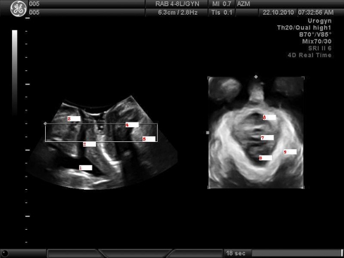
Methods: In this prospective observational cohort study, 245 women undergoing anterior colporrhaphy were invited for a 2-year follow-up visit consisting of a questionnaire, physical examination, and translabial 3D ultrasonography. Women with and without recurrent cystocele were compared to identify recurrence risk factors.
Results: Of the 245 women, 156 agreed to the follow-up visit (63.7%). Objective recurrence rate was 80 of 156 (51.3%). Seventeen of the 156 (10.9%) reported subjective recurrence. Risk factors for anatomical recurrence were complete avulsion of puborectalis muscle (OR, 2.4; 95% CI, 1.3, 4.7), advanced preoperative stage (OR, 2.0; 95% CI, 1.0, 4.1), family history of prolapse (OR, 2.4; 95% CI, 1.2, 4.9), and sacrospinous fixation (OR, 6.5; 95% CI, 2.0, 21.2).
Conclusions: Risk factors for anatomical cystocele recurrence after anterior colporrhaphy were complete avulsion of puborectalis muscle, advanced preoperative stage, family history of prolapse, and sacrospinous fixation.
Figures
References
-
- Miedel A, Tegerstedt G, Morlin B, Hammarstrom M. A 5-year prospective follow-up study of vaginal surgery for pelvic organ prolapse. Int Urogynecol J Pelvic Floor Dysfunct. 2008;9:1593–1601. - PubMed
MeSH terms
LinkOut - more resources
Full Text Sources
