. 2012 Aug;37(4):549-57.
doi: 10.1007/s00261-011-9790-1.

Deep infiltrating endometriosis of the bowel: MR imaging as a method to predict muscular invasion

Affiliations

Deep infiltrating endometriosis of the bowel: MR imaging as a method to predict muscular invasion

Milou P H Busard et al. Abdom Imaging. 2012 Aug.

Abstract

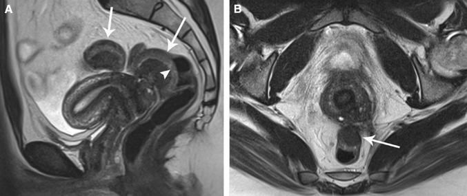
Purpose: To evaluate magnetic resonance (MR) imaging morphologic- and signal intensity abnormalities of deep infiltrating endometriosis (DIE) of the bowel wall and to assess its value in predicting depth and extent of bowel wall infiltration.

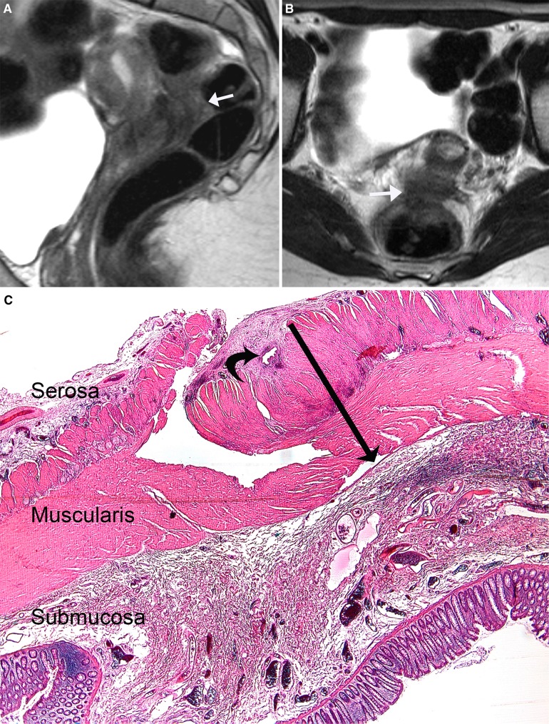
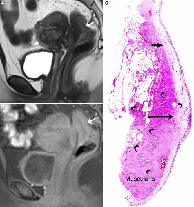
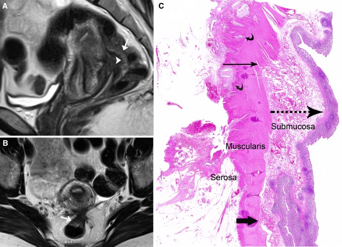
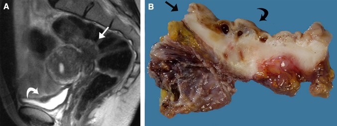
Materials and methods: This single-center study was performed in a tertiary referral center for endometriosis. All patients (n = 28) who underwent segmental bowel resection (2004-2010) were retrospectively studied. MR images were analyzed by two experienced readers independently (number of lesions, location, size, signal intensity, and depth of bowel wall infiltration) and this was correlated with histopathology.

Results: The sensitivity, specificity, positive and negative predictive values, and accuracy for diagnosis of endometriosis infiltrating the muscular layer of the bowel were 100%, 75%, 96%, 100%, and 96%, respectively. The inter-rater agreement was 0.84. "Fan shaped" configurations with hypointensity on T2- and T1-weighted imaging were characteristic for thickening of indigenous smooth muscle and smooth muscle hyperplasia at histopathology, as a consequence of infiltration by endometriosis. Thickening of the (sub)mucosa corresponded to edema with or without infiltration of endometriosis.

Conclusion: MR imaging at 1.5 Tesla is useful to predict muscular infiltration of the bowel in endometriosis, whereas it is of limited value in diagnosis of (sub)mucosal infiltration.


Figures

Fig. 1
Fig. 1
A 31-year-old woman who presented with dyschezia and hematochezia. Segmental bowel resection was performed, as hormonal therapy did not decrease pain symptoms in this patient. A and B Sagittal and axial T2-weighted images (6000–10000/137) show a lesion with isointense to slightly hyperintense signal compared to muscle in the posterior fornix that is infiltrating the bowel wall. The lesion was scored on MR imaging as infiltrating the serosa of the bowel wall without prominent thickening of the muscularis. C Histopathology shows endometriosis of the bowel serosa with fibrosis (curved black arrow) and minor local involvement of the muscular layer at this site. The indigenous smooth muscle is locally only minimally thickened (black arrow)
Fig. 2
Fig. 2
A 30-year-old woman known with endometriosis previously presented elsewhere with complete bowel obstruction, due to endometriosis infiltrating the rectosigmoid. Colostomy was performed before referral to our hospital. Thereafter, a segmental bowel resection was performed in our hospital. At histopathology, the endometriotic lesion was diagnosed as infiltrating the muscular and submucosal layers of the bowel wall. A Sagittal T2-weighted image (6000–10000/137) shows “fan shaped” configuration with isointense signal compared to muscle. B Sagittal T1-weighted image (740–790/19) shows isointensity of the lesion. C Histopathology shows endometriosis infiltrating the muscular and submucosal layer of the bowel wall (curved black arrows). At the site of endometriosis, thickening of indigenous smooth muscle is seen (black arrow) compared to the thickness of the indigenous smooth muscle in another area (thick black arrow).
Fig. 3
Fig. 3
A 36-year-old woman presented with progressive dyschezia and pencil-like stools. At colonoscopy, 10 cm from the anal canal swollen, edematous mucosa was seen with small submucosal bleedings. A and B Sagittal and axial T2-weighted images (6000–10000/137) show “fan shaped” configuration (white arrow) located at the torus uterinus with isointense signal compared to muscle and slightly high signal intensity at the luminal side of the bowel wall (arrowhead). C Histopathology shows endometriosis infiltrating the muscular layer of the bowel wall (curved black arrows). The image shows thickening of the muscular layer of the bowel wall at the site of endometriosis (black arrow), compared to a site without infiltration of endometriosis (thick black arrow). Furthermore “non-specific” inflammation with thickening of the submucosa (dotted arrow) is shown.
Fig. 4
Fig. 4
A 35-year-old woman, known with endometriosis previously presented with dyschezia, hematochezia, and pencil-like stools. Segmental bowel resection was performed and histopathology showed the lesion infiltrated the muscular and submucosal layer of the bowel wall. Non-specific inflammation was found with thickening of the submucosa. A Sagittal T2-weighted image (6000–10000/137) shows lesion infiltrating the rectosigmoid with isointense signal compared to muscle and slightly high signal intensity at the luminal side of the bowel wall (white arrow). Furthermore endometriosis is also found infiltrating the bladder wall (white curved arrow). B Macroscopic appearance shows bowel wall (black arrow corresponding to the rectum) with endometriosis infiltrating the muscular and submucosal layers (black curved arrow).
Fig. 5
Fig. 5
A 28-year-old woman diagnosed with two DIE lesions infiltrating the rectosigmoid and sigmoid. A and B Sagittal and axial T2-weighted images (6000–10000/137) show two lesions depicting “fan shaped” configurations (arrows), demonstrating isointense signal compared to muscle. In addition, hyperintense signal compared to muscle is found at the luminal side of the bowel wall (arrowhead).

References

    1. Balleyguier C, Chapron C, Dubuisson JB, et al. Comparison of magnetic resonance imaging and transvaginal ultrasonography in diagnosing bladder endometriosis. J Am Assoc Gynecol Laparosc. 2002;9(1):15–23. doi: 10.1016/S1074-3804(05)60099-0. - DOI - PubMed
    1. Bazot M, Darai E, Hourani R, et al. Deep pelvic endometriosis: MR imaging for diagnosis and prediction of extension of disease. Radiology. 2004;232(2):379–389. doi: 10.1148/radiol.2322030762. - DOI - PubMed
    1. Bazot M, Thomassin I, Hourani R, Cortez A, Darai E. Diagnostic accuracy of transvaginal sonography for deep pelvic endometriosis. Ultrasound Obstet Gynecol. 2004;24(2):180–185. doi: 10.1002/uog.1108. - DOI - PubMed
    1. Bazot M, Lafont C, Rouzier R, et al. Diagnostic accuracy of physical examination, transvaginal sonography, rectal endoscopic sonography, and magnetic resonance imaging to diagnose deep infiltrating endometriosis. Fertil Steril. 2009;92(6):1825–1833. doi: 10.1016/j.fertnstert.2008.09.005. - DOI - PubMed
    1. Bazot M, Gasner A, Lafont C, Ballester M, Darai E (2011) Deep pelvic endometriosis: limited additional diagnostic value of postcontrast in comparison with conventional MR images. Eur J Radiol. doi:0.1016/j.ejrad.2010.12.006 - PubMed