Comparative Study
. 2011 Oct 3;8(5):1582-91.
doi: 10.1021/mp200171d. Epub 2011 Aug 24.

Utilizing cell-matrix interactions to modulate gene transfer to stem cells inside hyaluronic acid hydrogels

Affiliations
Comparative Study

Utilizing cell-matrix interactions to modulate gene transfer to stem cells inside hyaluronic acid hydrogels

Shiva Gojgini et al. Mol Pharm. .

Abstract

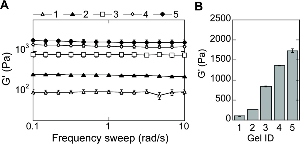
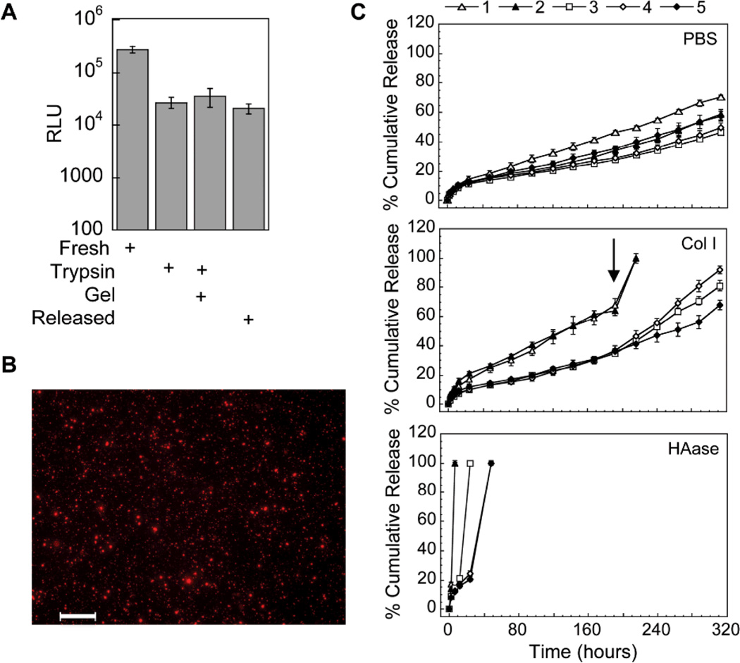
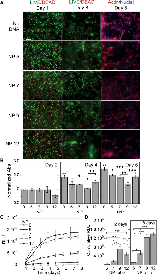
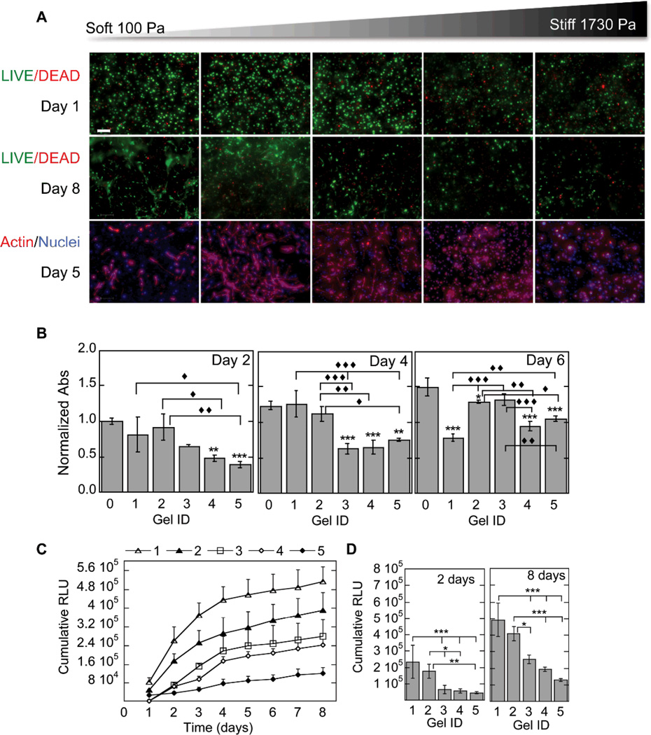
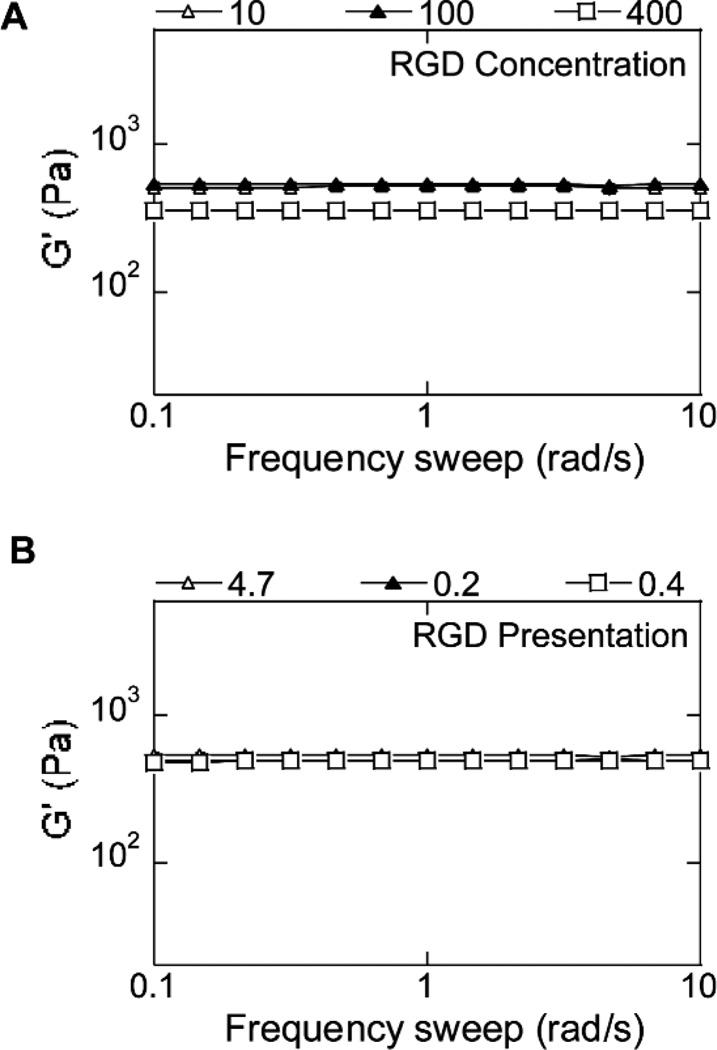
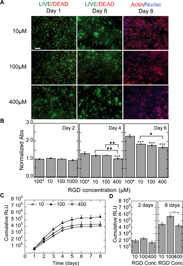
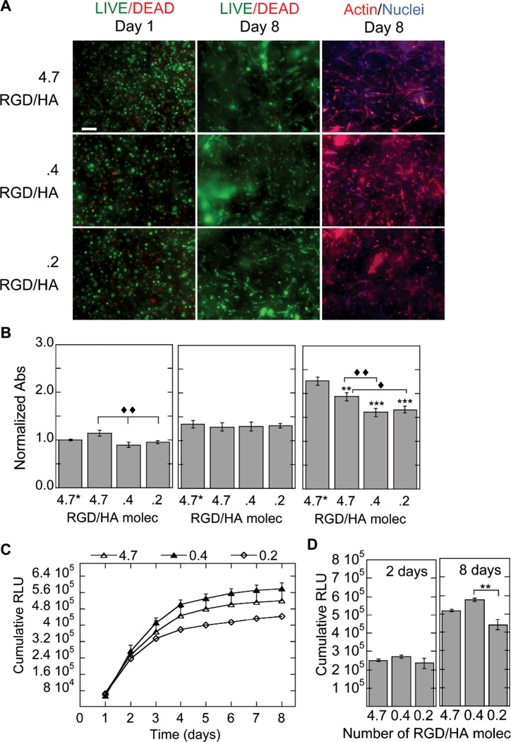
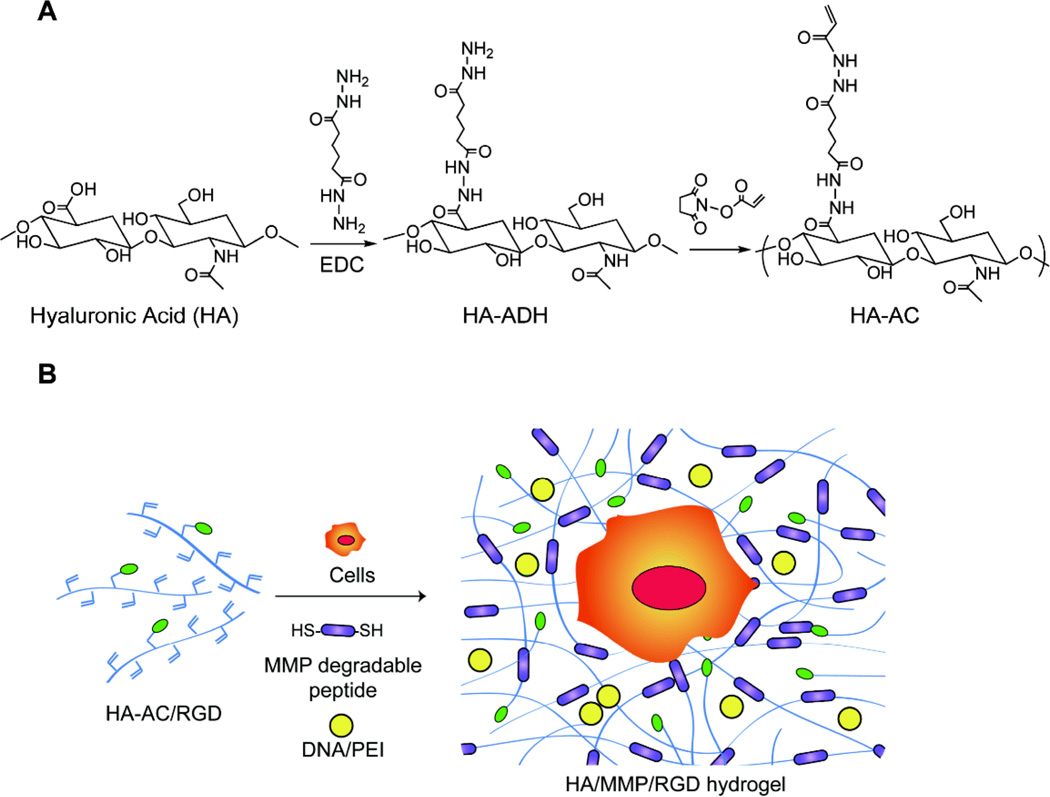
The effective delivery of DNA locally would increase the applicability of gene therapy in tissue regeneration, where diseased tissue is to be repaired in situ. One promising approach is to use hydrogel scaffolds to encapsulate and deliver plasmid DNA in the form of nanoparticles to the diseased tissue, so that cells infiltrating the scaffold are transfected to induce regeneration. This study focuses on the design of a DNA nanoparticle-loaded hydrogel scaffold. In particular, this study focuses on understanding how cell-matrix interactions affect gene transfer to adult stem cells cultured inside matrix metalloproteinase (MMP) degradable hyaluronic acid (HA) hydrogel scaffolds. HA was cross-linked to form a hydrogel material using a MMP degradable peptide and Michael addition chemistry. Gene transfer inside these hydrogel materials was assessed as a function of polyplex nitrogen to phosphate ratio (N/P = 5 to 12), matrix stiffness (100-1700 Pa), RGD (Arg-Gly-Asp) concentration (10-400 μM), and RGD presentation (0.2-4.7 RGDs per HA molecule). All variables were found to affect gene transfer to mouse mensenchymal stem cells culture inside the DNA loaded hydrogels. As expected, higher N/P ratios lead to higher gene transfer efficiency but also higher toxicity; softer hydrogels resulted in higher transgene expression than stiffer hydrogels, and an intermediate RGD concentration and RGD clustering resulted in higher transgene expression. We believe that the knowledge gained through this in vitro model can be utilized to design better scaffold-mediated gene delivery for local gene therapy.


Figures

Figure 1
Figure 1
Hydrogel mechanical properties. The mechanical properties of the hydrogels were determined using plate-to-plate rheometry Storage (A) and average (B) modulus over a frequency range of 0.1 to 10 rad/s at a constant strain of .03 are shown for increasingly stiff hydrogels (Gel ID 1 < 2 < 3 < 4 < 5).
Figure 2
Figure 2
Polyplex activity, distribution inside hydrogel scaffolds and release. (A) Activity of the entrapped polyplexes was determined through the release of the polyplexes post hydrogel formation using trypsin and a subsequent bolus transfection with the released polyplexes. The gene transfer of the released polyplexes was compared to fresh polyplexes with trypsin added and fresh polyplexes with gel degradation products added. (B) DNA/PEI polyplexes were stained with ethidium bromide post hydrogel formation and imaged with a fluorescence microscope equipped with z-stack capability. Scale bar = 100µm. (C) DNA release was determined using radiolabeled DNA. DNA/PEI loaded hydrogels were incubated in different release solutions and at predetermined time points samples were gathered and analyzed for radioactivity using a scintillation counter. At the final day of the release assay the hydrogel was fully degraded with trypsin and the final activity measured. Data is plotted as the % cumulative release.
Figure 3
Figure 3
Gene transfer as a function of N/P ratio. The effect of N/P ratio on transgene expression was studied for cells cultured inside MMP degradable HA hydrogels. For these studies a 3% hydrogel with an r ratio of 0.3 was used. The cell viability, ability of the cells to spread and the metabolic activity of the cells were studied using the LIVE/DEAD assay, phalloidin staining (A) and MTT assay (B). Gene expression was determined over time using a reporter plasmid, gaussia luciferase, which is secreted by the cell when expressed (C). The cumulative expression at days 2 and 8 is plotted for ease of comparison (D). Statistical significance was determined using multiple comparisons and either the Dunnett or the Tukey multiple comparison’s tests. The symbol ** indicates statistical significance at the level of 0.01 between the indicated condition and the corresponding no DNA control in (B) or between the indicated conditions in (D). The symbols ♦, ♦♦, ♦♦♦ indicate statistical significance at the level of 0.05, 0.01 and 0.001 between the indicated conditions in (B). N/P = 0 represents the condition with no DNA polyplexes added to the hydrogel. Scale bar = 100µm.
Figure 4
Figure 4
Gene transfer as a function of hydrogel stiffness. The effect of hydrogel stiffness on the ability of cells seeded inside the hydrogel to become transfected was studied for hydrogels with storage modulus ranging from 100 Pa to 1730 Pa. The cell viability, ability of the cells to spread and the metabolic activity of the cells was studied using the LIVE/DEAD assay, phalloidin staining (A) and MTT assay (B). None of the cell stiffness resulted in lower cellular viability. However, cell spreading was inhibited for stiffer hydrogels. Gene expression was determined over time using a reporter plasmid, gaussia luciferase, which is secreted by the cell when expressed (C). The cumulative expression at days 2 and 8 is plotted for ease of comparison (D). Matrix stiffness influenced transgene expression. The numbers 1–5 represent different hydrogel stiffness. 1 = 100 Pa, 2 = 260 Pa, 3 = 839 Pa, 4 = 1360 Pa, 5 = 1730 Pa. Statistical significance was determined using multiple comparisons and either the Dunnett or the Tukey multiple comparison’s tests. The symbols ** and *** indicate statistical significance at the level of 0.05, 0.01 and 0.001 between the indicated condition and the corresponding no DNA control in (B) or between the indicated conditions in (D). The symbols ♦, ♦♦, ♦♦♦ indicate statistical significance at the level of 0.05, 0.01 and 0.001 between the indicated conditions in (B). Gel ID 0 represents the condition with no DNA polyplexes added to the hydrogel. Scale bar = 100µm.
Figure 5
Figure 5
Hydrogel mechanical properties for hydrogels with different RGD concentrations and presentations. The mechanical properties of the hydrogels were determined using plate-to-plate rheometry Storage (A, B)modulus over a frequency range of 0.1 to 10 rad/s at a constant strain of 0.03 are shown for hydrogels with various RGD concentrations and presentations, respectively. RGD presentation is displayed as number of RGD/HA molecule with 4.7 RGD/HA being the most clustered condition and .2 RGD/HA being the least clustered/homogeneously distributed condition.
Figure 6
Figure 6
Gene transfer as a function of RGD concentration. The effect of RGD concentration on the ability of cells seeded inside the hydrogel to become transfected was studied for hydrogels with RGD ranging from 10 µM to 400 µM. The cell viability, ability of the cells to spread and the metabolic activity of the cells was studied using the LIVE/DEAD assay, phalloidin staining (A) and MTT assay (B). Gene expression was determined over time using a reporter plasmid, gaussia luciferase, which is secreted by the cell when expressed (C). The cumulative expression at days 2 and 8 is plotted for ease of comparison (D). Different RGD concentration influenced transgene expression. Statistical significance was determined using multiple comparisons and either the Dunnett or the Tukey multiple comparison’s tests. The symbol *** indicates statistical significance at the level of 0.001 between the indicated condition and the no DNA control in (B) or between the indicated conditions in (D). The symbols ♦, ♦♦, ♦♦♦ indicate statistical significance at the level of 0.05, 0.01 and 0.001 between the indicated conditions in (B). 100* represents the condition with no DNA polyplexes added to the hydrogel. Scale bar = 100µm.
Figure 7
Figure 7
Gene transfer as a function of RGD presentation. The effect of RGD presentation on the ability of cells seeded inside the hydrogel to become transfected was studied for hydrogels with 100 µM RGD displayed either homogeneously (100% HA-RGD, .2 RGD/HA molecule) or as RGD clusters (52% to 4.3% HA-RGD, .4 and 4.7 HA/RGD molecule, respectively). RGD clustering is achieved by reacting different amounts of HA-AC with the same amount of RGD and then mixing the resulting HA-RGD with unmodified HA. The cell viability, ability to of the cells to spread and the metabolic activity of the cells were studied using the LIVE/DEAD assay, phalloidin staining (A) and MTT assay (B). Gene expression was determined over time using a reporter plasmid, gaussia luciferase, which is secreted by the cell when expressed (C). The cumulative expression at days 2 and 8 is plotted for ease of comparison (D). RGD presentation influenced transgene expression. Statistical significance was determined using multiple comparisons and either the Dunnett or the Tukey multiple comparison’s tests. The symbols ** and *** indicates statistical significance at the level of 0.01 and 0.001 between the indicated condition and the corresponding no DNA control in (B) and between the indicated conditions in (D). The symbols ♦, ♦♦ indicate statistical significance at the level of 0.05 and 0.01 between the indicated conditions in (B). 4.7* represents the condition with no DNA polyplexes added to the hydrogel. Scale bar = 100µm.
Scheme 1
Scheme 1
chematic of HA modification and hydrogel formation. (A) HA-acrylate synthesis is a two-step process first reacting HA with ADH and then using the pendant hydrazide to react with NHS-acrylate. (B) Schematic of DNA-loaded hydrogel formation. Liquid HA-AC is first modified with RGD peptides using Michael type addition. HA-RGD is then crosslinked using an MMP degradable peptide in the presence of DNA/PEI polyplexes.

References

    1. Kleinman HK, Philp D, Hoffman MP. Role of the extracellular matrix in morphogenesis. Curr Opin Biotechnol. 2003;14(5):526–532. - PubMed
    1. Bottaro DP, Liebmann-Vinson A, Heidaran MA. Molecular signaling in bioengineered tissue microenvironments. Ann N Y Acad Sci. 2002;961:143–153. - PubMed
    1. Fraser JR, Laurent TC, Laurent UB. Hyaluronan: its nature, distribution, functions and turnover. J Intern Med. 1997;242(1):27–33. - PubMed
    1. Leach JB, Bivens KA, Patrick CW, Schmidt CE. Photocrosslinked hyaluronic acid hydrogels: Natural, biodegradable tissue engineering scaffolds. Biotechnology and Bioengineering. 2003;82(5):578–589. - PubMed
    1. Park YD, Tirelli N, Hubbell JA. Photopolymerized hyaluronic acid-based hydrogels and interpenetrating networks. Biomaterials. 2003;24(6):893–900. - PubMed

Publication types

MeSH terms