. 2011 Aug 7;135(5):055106.
doi: 10.1063/1.3623418.

Two-dimensional infrared spectroscopy of azido-nicotinamide adenine dinucleotide in water

Affiliations

Two-dimensional infrared spectroscopy of azido-nicotinamide adenine dinucleotide in water

Samrat Dutta et al. J Chem Phys. .

Abstract

Mid-IR active analogs of enzyme cofactors have the potential to be important spectroscopic reporters of enzyme active site dynamics. Azido-nicotinamide adenine dinucleotide (NAD(+)), which has been recently synthesized in our laboratory, is a mid-IR active analog of NAD(+), a ubiquitous redox cofactor in biology. In this study, we measure the frequency-frequency time correlation function for the antisymmetric stretching vibration of the azido group of azido-NAD(+) in water. Our results are consistent with previous studies of pseudohalides in water. We conclude that azido-NAD(+) is sensitive to local environmental fluctuations, which, in water, are dominated by hydrogen-bond dynamics of the water molecules around the probe. Our results demonstrate the potential of azido-NAD(+) as a vibrational probe and illustrate the potential of substituted NAD(+)-analogs as reporters of local structural dynamics that could be used for studies of protein dynamics in NAD-dependent enzymes.


Figures

Figure 1
Figure 1
Structures for nicotinamide adenine dinucleotide (NAD+), left, and azido-nicotinamide adenine dinucleotide (azido-NAD+), right.
Figure 2
Figure 2
Spectral slices of the dispersed pump-probe decay for delay times T = 500 fs (red), 1 ps (blue), and 2 ps (black).
Figure 3
Figure 3
Decay of the excited-state absorption contribution to the dispersed pump-probe spectrum as a function of the pump-probe delay. The transient is fit to a single exponential decay with a time constant of 1.0 ps.
Figure 4
Figure 4
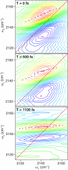
2D-IR spectra of azido-NAD+ for population times T = 0 fs, 500 fs, and 1.1 ps. The open circles mark the center lines and the solid red lines are the linear fits to the centerlines that we use to determine the CLS.
Figure 5
Figure 5
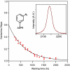
CLS vs waiting time (open circles) along with the exponential fit to the CLS decay. The inset compares the measured (red) and calculated (black) antisymmetric stretch absorption spectrum of the azido-NAD+ chromophore. The structure of Azido-NAD+ is also shown (ADPR: adenine pyrophosphate).

Similar articles

Cited by

References

    1. Bandaria J., Cheatum C. M., and Kohen A., J. Am. Chem. Soc. 131(29), 10151 (2009).10.1021/ja902120t - DOI - PMC - PubMed
    1. Wang L., Goodey N. M., Benkovic S. J., and Kohen A., Philos. Trans. R. Soc. London, Ser. B 361(1472), 1307 (2006).10.1098/rstb.2006.1871 - DOI - PMC - PubMed
    1. Oyeyemi O. A., Sours K. M., Lee T., Resing K. A., Ahn N. G., and Klinman J. P., Proc. Natl. Acad. Sci. U.S.A. 107(22), 10074 (2010).10.1073/pnas.1003678107 - DOI - PMC - PubMed
    1. Klinman J. P., Chem. Phys. Lett. 471(4–6), 179 (2009).10.1016/j.cplett.2009.01.038 - DOI - PMC - PubMed
    1. Hay S., Pudney C. R., McGrory T. A., Pang J. Y., Sutcliffe M. J., and Scrutton N. S., Angew. Chem., Int. Ed. 48(8), 1452 (2009).10.1002/anie.200805502 - DOI - PubMed

Publication types