Review
. 2011 Jul;133(7):071010.
doi: 10.1115/1.4004493.

Spinal facet joint biomechanics and mechanotransduction in normal, injury and degenerative conditions

Affiliations
Review

Spinal facet joint biomechanics and mechanotransduction in normal, injury and degenerative conditions

Nicolas V Jaumard et al. J Biomech Eng. 2011 Jul.

Abstract

The facet joint is a crucial anatomic region of the spine owing to its biomechanical role in facilitating articulation of the vertebrae of the spinal column. It is a diarthrodial joint with opposing articular cartilage surfaces that provide a low friction environment and a ligamentous capsule that encloses the joint space. Together with the disc, the bilateral facet joints transfer loads and guide and constrain motions in the spine due to their geometry and mechanical function. Although a great deal of research has focused on defining the biomechanics of the spine and the form and function of the disc, the facet joint has only recently become the focus of experimental, computational and clinical studies. This mechanical behavior ensures the normal health and function of the spine during physiologic loading but can also lead to its dysfunction when the tissues of the facet joint are altered either by injury, degeneration or as a result of surgical modification of the spine. The anatomical, biomechanical and physiological characteristics of the facet joints in the cervical and lumbar spines have become the focus of increased attention recently with the advent of surgical procedures of the spine, such as disc repair and replacement, which may impact facet responses. Accordingly, this review summarizes the relevant anatomy and biomechanics of the facet joint and the individual tissues that comprise it. In order to better understand the physiological implications of tissue loading in all conditions, a review of mechanotransduction pathways in the cartilage, ligament and bone is also presented ranging from the tissue-level scale to cellular modifications. With this context, experimental studies are summarized as they relate to the most common modifications that alter the biomechanics and health of the spine-injury and degeneration. In addition, many computational and finite element models have been developed that enable more-detailed and specific investigations of the facet joint and its tissues than are provided by experimental approaches and also that expand their utility for the field of biomechanics. These are also reviewed to provide a more complete summary of the current knowledge of facet joint mechanics. Overall, the goal of this review is to present a comprehensive review of the breadth and depth of knowledge regarding the mechanical and adaptive responses of the facet joint and its tissues across a variety of relevant size scales.


Figures

Fig. 1
Fig. 1
Lateral view of a cervical (a) and axial view of a lumbar (b) vertebra showing the overall anatomy and the facet joints, articulations, and orientation relative to its angle with each of the axial plane (β) and of the sagittal plane (α)
Fig. 2
Fig. 2
Schematic drawings of the facet joint and the primary tissues that compose it, as well as the cartilage and menisci of the facet articulation. The blowup illustrates the different zones of the articular cartilage layer with the collagen fibers and chondrocytes orientations through its depth. A cut through of the facet joint (A-A) is also drawn to show the elliptically-shaped inter-articular surfaces with the cartilage surface on the inferior facet, the synovium, and meniscoids. Adapted collectively from Martin et al., 1998, Pierce et al., 2009, and Bogduk and Engel, 1984 [48,49,73].
Fig. 3
Fig. 3
Representative data quantifying the spinal rotations and pressure responses in the facet of a multisegment (C2-T1) cadaveric cervical spine during a range of bending moments applied in continuous flexion-extension. The pressure response in the C5-C6 facet joint increases with applied extension as contact is developed in the articulating facets, but exhibits a different pattern than the rotation angle. In contrast, during flexion, when the joint opens up there is no pressure detected.
Fig. 4
Fig. 4
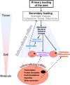
Schematic representation of the generalized processes of mechanotransduction in synovial joints, across the scales ranging from tissue to molecule
Fig. 5
Fig. 5
(a) Lateral radiographic of a healthy spine, indicating a healthy C5-C6 facet joint. A tip-mounted transducer has been inserted in the superior articular facet to measure the contact pressure developed in the facet joint during experimental studies inducing spinal bending. (b) A photograph of the facet surface of an exposed C5 facet from a 65 year old male donor demonstrating hallmarks of a degenerated articular surface: fissure (single arrow) and eroded cartilage area (double arrow).
Fig. 6
Fig. 6
Schematic illustrating the multifaceted approach to understanding the form, function and physiological balance of mechanisms in the facet joint. A variety of experimental and computational techniques are needed to complement the existing knowledge of the factors that directly and indirectly affect the physiologic performance of the facet joint, as well as its function in health, injury, aging and spinal interventions.

Similar articles

Cited by

References

    1. Adams, M. A. and Hutton, W.C. , 1983, “The Mechanical Function of Lumbar Apophyseal Joints,” Spine, 8, pp. 327–330.10.1097/00007632-198304000-00017 - DOI - PubMed
    1. Yang, K. H. and King, A. I. , 1984, “Mechanism of Facet Load Transmission as a Hypothesis for Low-Back Pain,” Spine, 9(6), pp. 557–565.10.1097/00007632-198409000-00005 - DOI - PubMed
    1. Schendel, M. J. , Dekutoski, M. B. , Ogilvie, J. W. , Olsewski, J. M. , and Wallace, L. J. , 1995, “Kinematics of The Canine Lumbar Intervertebral Joint. An in Vivo Study Before and After Adjacent Instrumentation,” Spine, 20(23), pp. 2555–2564.10.1097/00007632-199512000-00015 - DOI - PubMed
    1. Pal, G. P. and Routal, R. V. , 1986, “A Study of Weight Transmission Through the Cervical and Upper Thoracic Regions of the Vertebral Column in Man,” J. Anat., 148, pp. 245–261. - PMC - PubMed
    1. Pal, G. P. and Routal, R. V. , 1987, “Transmission of Weight Through the Lower Thoracic and Lumbar Regions of the Vertebral Column in Man,” J. Anat., 152, pp. 93–105. - PMC - PubMed

Publication types

MeSH terms