

Metabolic alteration of urinary steroids in pre- and post-menopausal women, and men with papillary thyroid carcinoma
- PMID: 21824401
- PMCID: PMC3199870
- DOI: 10.1186/1471-2407-11-342
Metabolic alteration of urinary steroids in pre- and post-menopausal women, and men with papillary thyroid carcinoma
Abstract
Background: To evaluate the metabolic changes in urinary steroids in pre- and post-menopausal women and men with papillary thyroid carcinoma (PTC).
Methods: Quantitative steroid profiling combined with gas chromatography-mass spectrometry was used to measure the urinary concentrations of 84 steroids in both pre- (n = 21, age: 36.95 ± 7.19 yr) and post-menopausal female (n = 19, age: 52.79 ± 7.66 yr), and male (n = 16, age: 41.88 ± 8.48 yr) patients with PTC. After comparing the quantitative data of the patients with their corresponding controls (pre-menopause women: n = 24, age: 33.21 ± 10.48 yr, post-menopause women: n = 16, age: 49.67 ± 8.94 yr, male: n = 20, age: 42.75 ± 4.22 yr), the levels of steroids in the patients were normalized to the mean concentration of the controls to exclude gender and menopausal variations.
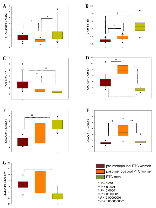
Results: Many urinary steroids were up-regulated in all PTC patients compared to the controls. Among them, the levels of three active androgens, androstenedione, androstenediol and 16α-hydroxy DHEA, were significantly higher in the pre-menopausal women and men with PTC. The corticoid levels were increased slightly in the PTC men, while progestins were not altered in the post-menopausal PTC women. Estrogens were up-regulated in all PTC patients but 2-hydroxyestrone and 2-hydroxy-17β-estradiol were remarkably changed in both pre-menopausal women and men with PTC. For both menopausal and gender differences, the 2-hydroxylation, 4-hydroxylation, 2-methoxylation, and 4-methoxylation of estrogens and 16α-hydroxylation of DHEA were differentiated between pre- and post-menopausal PTC women (P < 0.001). In particular, the metabolic ratio of 2-hydroxyestrone to 2-hydroxy-17β-estradiol, which could reveal the enzyme activity of 17β-hydroxysteroid dehydrogenase, showed gender differences in PTC patients (P < 1 × 10⁻⁷).
Conclusions: These results are expected be helpful for better understanding the pathogenic differences in PTC according to gender and menopausal conditions.
Figures

Similar articles
-
Delta-5-androstenediol and its sulphate in serum and urine of normal adults and patients with endocrine diseases.Clin Endocrinol (Oxf). 1993 Oct;39(4):475-82. doi: 10.1111/j.1365-2265.1993.tb02396.x. Clin Endocrinol (Oxf). 1993. PMID: 8287575
-
Serum concentrations of selected endogenous estrogen and estrogen metabolites in pre- and post-menopausal Chinese women with osteoarthritis.J Endocrinol Invest. 2010 Oct;33(9):644-9. doi: 10.1007/BF03346664. Epub 2010 Mar 25. J Endocrinol Invest. 2010. PMID: 20339312
-
Fat tissue: a steroid reservoir and site of steroid metabolism.J Clin Endocrinol Metab. 1985 Sep;61(3):564-70. doi: 10.1210/jcem-61-3-564. J Clin Endocrinol Metab. 1985. PMID: 3160722
-
All sex steroids are made intracellularly in peripheral tissues by the mechanisms of intracrinology after menopause.J Steroid Biochem Mol Biol. 2015 Jan;145:133-8. doi: 10.1016/j.jsbmb.2014.06.001. Epub 2014 Jun 9. J Steroid Biochem Mol Biol. 2015. PMID: 24923731 Review.
-
Plasma levels and secretion rate of steroids with anabolic activity in man.Environ Qual Saf Suppl. 1976;(5):171-80. Environ Qual Saf Suppl. 1976. PMID: 133798 Review.
Cited by
-
Oncometabolites as biomarkers in thyroid cancer: a systematic review.Cancer Manag Res. 2019 Feb 25;11:1829-1841. doi: 10.2147/CMAR.S188661. eCollection 2019. Cancer Manag Res. 2019. PMID: 30881111 Free PMC article. Review.
-
Metabolic profiling of cholesterol and sex steroid hormones to monitor urological diseases.Endocr Relat Cancer. 2016 Oct;23(10):R455-67. doi: 10.1530/ERC-16-0285. Endocr Relat Cancer. 2016. PMID: 27580660 Free PMC article. Review.
-
Predictors of body composition and body energy changes in response to chronic overfeeding.Int J Obes (Lond). 2014 Feb;38(2):236-42. doi: 10.1038/ijo.2013.77. Epub 2013 May 20. Int J Obes (Lond). 2014. PMID: 23736367 Free PMC article.
-
A review of complex hormone regulation in thyroid cancer: novel insights beyond the hypothalamus-pituitary-thyroid axis.Front Endocrinol (Lausanne). 2024 Jul 22;15:1419913. doi: 10.3389/fendo.2024.1419913. eCollection 2024. Front Endocrinol (Lausanne). 2024. PMID: 39104813 Free PMC article. Review.
-
FKBP51 promotes migration and invasion of papillary thyroid carcinoma through NF-κB-dependent epithelial-to-mesenchymal transition.Oncol Lett. 2018 Dec;16(6):7020-7028. doi: 10.3892/ol.2018.9517. Epub 2018 Sep 27. Oncol Lett. 2018. PMID: 30546435 Free PMC article.
References
-
- Sakoda LC, Horn-Ross PL. Reproductive and menstrual history and papillary thyroid cancer risk: the San Francisco Bay area thyroid cancer study. Cancer Epidemiol Biomarks Prev. 2002;11:51–57. - PubMed
Publication types
MeSH terms
Substances
LinkOut - more resources
Full Text Sources
Medical
Molecular Biology Databases
