Review
. 2011 Nov;30(6):405-30.
doi: 10.1016/j.preteyeres.2011.07.002. Epub 2011 Jul 29.

The functional cycle of visual arrestins in photoreceptor cells

Affiliations
Review

The functional cycle of visual arrestins in photoreceptor cells

Vsevolod V Gurevich et al. Prog Retin Eye Res. 2011 Nov.

Abstract

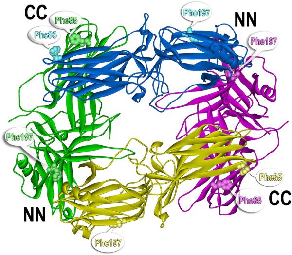
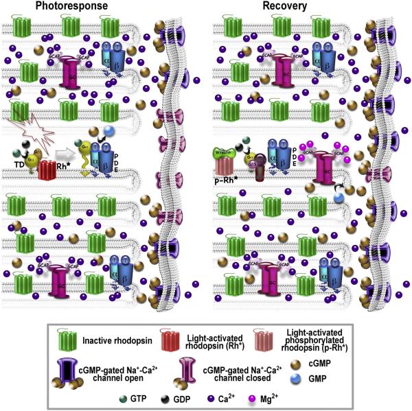
Visual arrestin-1 plays a key role in the rapid and reproducible shutoff of rhodopsin signaling. Its highly selective binding to light-activated phosphorylated rhodopsin is an integral part of the functional perfection of rod photoreceptors. Structure-function studies revealed key elements of the sophisticated molecular mechanism ensuring arrestin-1 selectivity and paved the way to the targeted manipulation of the arrestin-1 molecule to design mutants that can compensate for congenital defects in rhodopsin phosphorylation. Arrestin-1 self-association and light-dependent translocation in photoreceptor cells work together to keep a constant supply of active rhodopsin-binding arrestin-1 monomer in the outer segment. Recent discoveries of arrestin-1 interaction with other signaling proteins suggest that it is a much more versatile signaling regulator than previously thought, affecting the function of the synaptic terminals and rod survival. Elucidation of the fine molecular mechanisms of arrestin-1 interactions with rhodopsin and other binding partners is necessary for the comprehensive understanding of rod function and for devising novel molecular tools and therapeutic approaches to the treatment of visual disorders.


Figures

Fig. 1
Fig. 1. Arrestin-1 selectivity for P-Rh* is achieved by a multi-step binding mechanism
A. Arrestin-1 demonstrates many times higher binding to P-Rh* than to inactive P-Rh or Rh*. Low-affinity interactions with dark P-Rh or Rh* are mediated by arrestin-1 elements that specifically recognize receptor-attached phosphates or the active state of rhodopsin, respectively. These elements act as phosphate and active conformation sensors, respectively. Only P-Rh* can engage both sites. Simultaneous activation of both sensors allows arrestin-1 transition from the basal (B) to the active high-affinity receptor-binding state (C). This transition involves the release of the arrestin C-tail (light brown), relatively small shifts in the relative positions of the N-domain (green) and C-domain (purple), significant rearrangement of flexible loops on the receptor-binding surface: “finger loop” (light green), “139 loop” (light blue), “344 loop” (dark blue), and “163 loop” (pink), and is limited by the length of the inter-domain hinge (yellow).
Fig. 2
Fig. 2. Critical inter-domain interactions disrupted during arrestin-1 activation by rhodopsin-attached phosphates
The arrestin-1 crystal structure (Hirsch et al., 1999) shows that the polar core, localized at the inter-domain interface, stabilizes the relative orientation of the two arrestin-1 domains. The polar core consists of five charged solvent-excluded residues (Asp30, Arg175, Asp296, Asp303, Arg382 shown as CPK models) that form a network of interactions and includes the salt bridge between Arg175 and Asp296 that serves as the main phosphate sensor. The C-tail folds back on the N-domain, where it participates in the three-element interaction with β-strand I and α-helix I. Bulky hydrophobic residues mediating this interaction are shown in yellow as CPK models. Two adjacent positive charges in β-strand I (Lys14 and Lys15, shown as CPK models) bind receptor-attached phosphates. Their engagement by phosphates and consequent change in orientation contributes to the destabilization of the three-element interaction, with subsequent release of the arrestin-1 C-tail (see Fig. 1). Destabilization of both interactions constitutes part of the arrestin-1 activation mechanism, facilitating its transition into a high-affinity rhodopsin-binding state.
Fig. 3
Fig. 3. Specific mutations in arrestin-1 pre-activate the phosphate sensor, yielding mutants with high affinity for active unphosphorylated Rh*
Targeted disruption of the salt bridge between Arg175 and Asp296 by charge reversal mutations R175E or D296R, as well as forced detachment of the arrestin-1 C-tail by triple alanine substitution of bulky hydrophobic residues that anchor it to the body of the N-domain (3A) yield partially activated arrestin-1 mutants with greatly increased binding to Rh*, and to a lesser extent, dark P-Rh. These data reveal the molecular mechanism of arrestin-1 activation and demonstrate the potential of targeted manipulation of arrestin-1 selectivity for different functional forms of rhodopsin.
Fig. 4
Fig. 4. Receptor specificity of arrestins is encoded in the structure of the receptor-binding surface
A. View of the receptor-binding surface. Four exposed residues on the concave side of the N-domain (Val52, Gly54, Lys55, Ile72) and six in the C-domain (Val244, Asn246, Lys257, Thr258, Gln265, Lys267) determine arrestin-1 preference for P-Rh*. Val90 (shown in red) makes the β-strand sandwich of the N-domain more rigid, ensuring strict receptor specificity of arrestin-1. Ser86 or Ala87 replace Val90 in non-visual arrestin-2 and -3, respectively, ensuring broad receptor specificity of these subtypes. The replacement of homologous arrestin-2 residues with those derived from arrestin-1 also fully reverses arrestin-2 receptor preference, yielding a mutant with very high binding to P-Rh* and low binding to other GPCRs (Vishnivetskiy et al., 2011). B. Side view of the arrestin-2 crystal structure (Han et al., 2001), where the elements that determine its receptor preference are shown in yellow, key receptor discriminator residues within these elements are shown in blue, and buried Ser86 is shown in red. In over 600 million years of arrestin evolution, very few residues (out of 20 possible) occupy the positions responsible for receptor preference (shown in quote bubbles) (Gurevich and Gurevich, 2010a).
Fig. 5
Fig. 5. Monomeric rhodopsin is the physiologically relevant target of GRK1 and arrestin-1
A,C. Rhodopsin in native disc membranes, as well as other GPCRs, can form dimers and other higher order oligomers. Although oligomeric forms of class A GPCRs tend to be very transient, with a sub-second half-life ((Fonseca and Lambert, 2009; Hern et al., 2010; Kasai et al., 2011; Lan et al., 2011); reviewed in (Gurevich and Gurevich, 2008b; Lambert, 2010)), their existence raises the question of whether monomers or dimers are biologically relevant partners of G proteins, GRKs, and arrestins. To determine the form of rhodopsin that is phosphorylated by GRK1 and binds arrestin, rhodopsin was reconstituted into nanodiscs as a monomer and its phosphorylation by purified GRK1 and the subsequent ability of monomeric P-Rh* to bind arrestin-1 were tested (Bayburt et al., 2011). The results showed that monomeric Rh* in nanodiscs is phosphorylated by GRK1 at least as efficiently as rhodopsin in disc membranes (B), and monomeric P-Rh* in nanodiscs bound arrestin-1 with physiologically relevant affinity (KD ~4 nM) and with the same 1:1 stoichiometry observed in vivo and in vitro in disc membranes (D). Along with evidence that monomeric Rh* efficiently activates transducin (Banerjee et al., 2008; Bayburt et al., 2007; Ernst et al., 2007; Whorton et al., 2008), these data demonstrate that the rhodopsin monomer is the functional unit at all steps of signaling and inactivation.
Fig. 6
Fig. 6. The molecular basis of the high Arrhenius energy of arrestin activation
A. The binding of WT arrestin-1 to P-Rh* has a high activation energy (Schleicher et al., 1989), and therefore can be inhibited by reduced temperature. Binding at low temperature is dramatically increased by the destabilization of the polar core (R175E mutation), forced detachment of the C-tail (3A mutation), or a combination of both (3A+R175E), indicating that these mutations significantly reduce the arrestin-1 activation energy. B. The binding of pre-activated mutants to Rh* is significantly lower at 20°C and especially at 0°C than at 37°C. Even the combination of 3A and R175E mutations, relieving both known conformational constraints in arrestin-1, yields arrestin-1 with four-fold lower binding to Rh* at 0°C than at 37°C. Thus, additional conformational constraints contribute to the activation energy of arrestin-1.
Fig. 7
Fig. 7. The structure of the solution tetramer of bovine arrestin-1
Inter-subunit distance measurements, mutagenesis, and the formation of inter-subunit disulfide bonds showed that the solution tetramer is dramatically different from the one observed in the crystal (Hanson et al., 2008a; Hanson et al., 2007c). The solution tetramer is virtually symmetrical, has a closed diamond shape with two N-domain-N-domain (NN) and two C-domain-C-domain (CC) inter-subunit interfaces. The receptor-binding concave sides of both domains are shielded by sister subunits in the tetramer and both possible dimers, explaining why only monomeric arrestin-1 can bind rhodopsin (Hanson et al., 2007c). Phe86 and Phe197 (shown as CPK models in all four subunits) play a key role in the stabilization of the NN and CC interfaces, respectively. Double alanine substitution in bovine (F86A, F197A) and mouse (F87A, F198A) arrestin-1 disrupts both interfaces, yielding constitutively monomeric mutants (Kim et al., 2011a). Thus, the shape of the solution tetramer is likely conserved between species.
Fig. 8
Fig. 8. The intricate shape of the rod cytoplasm and highly localized signaling suppress the variability of the single photon response
The biochemical signal amplification cascade Rh*-Td-PDE, and Rh* inactivation by GRK1 phosphorylation followed by arrestin-1 binding both involve stochastic processes, yet the single photon response in rods, measured as the light-induced change of current at the plasma membrane, shows much better reproducibility than expected. Theoretically, a large number of inactivation steps could serve as variability suppressor (Rieke and Baylor, 1998), but only if the contribution of all steps to Td and PDE activation is roughly equal. Since this is not the case, and as rhodopsin is likely inactivated in only four steps (three phosphorylations and arrestin-1 binding), other mechanisms must ensure high reproducibility of the response. A spatiotemporal mathematical model shows that the variability is suppressed by highly localized signaling initiated by a single Rh*.The biochemical changes occur within a narrow slice of the rod around the activated disc due to the complex shape of the rod cytoplasm that prevents signal spreading (Bisegna et al., 2008; Caruso et al., 2011). Photoresponse is “standardized” by the fact that PDE rapidly depletes cGMP in the vicinity of the disc containing active Rh*, so that PDE generated by the unusually long-lived Rh* has little substrate to hydrolyze, and therefore increases the variation less than it would in a well-stirred model. High cooperativity of the effect of cGMP concentration on channel opening probability further reduces the variability, ensuring that current suppression translates contiguous cGMP changes into a highly localized and essentially “all-or-nothing” channel response. The recovery phase is also highly localized. Moreover, channel closure results in an immediate local drop in Ca2+, which is replaced with the constantly present Mg2+ on GCAPs, converting them into activators of guanylate cyclase (GC) (Dizhoor et al., 2010). The ensuing rapid synthesis of cGMP replenishes its local concentration, opening the channels and returning the rod to the initial state. The activation of one rhodopsin by a single photon in dark-adapted rod suppresses 3-5% of dark current, which is equivalent to channel closure in the membrane surrounding 24-40 discs (out of ~800 in mouse rod). To make illustration of the local nature of the single photon response possible, each disc shown here represents multiple discs in the real OS. Key molecules involved in signaling and recovery are shown below the schematics.
Fig. 9
Fig. 9. Arrestin-1 interacts with all three kinases in the ASK1-MKK4-JNK3 signaling module
A, C. A nuclear exclusion assay demonstrates arrestin-1 interactions with JNK3 and ASK1. JNK3 (A) and ASK1 with an engineered nuclear localization sequence (NLS) (C) predominantly localize to the nucleus. Arrestin-1 has internal nuclear export signals that ensure its cytoplasmic localization. Co-expressed arrestin-1 relocalizes JNK3 and ASK1-NLS from the nucleus to the cytoplasm, similar to non-visual arrestin-2 and arrestin-3, as well as cone arrestin-4. Lower panels in A and C show the fraction of cells where more JNK3 (A) or ASK1-NLS (C) was localized in the cytoplasm than in the nucleus (C>N). Thus, arrestin-1 binds JNK3 and ASK1 (and that in complex the nuclear export signal of arrestin-1 is stronger than the NLS of JNK3 or ASK1). B. Co-immunoprecipitation shows arrestin-1 interaction with MKK4 in cells. Flag-tagged arrestins are immunoprecipitated by anti-Flag antibodies. Co-expressed HA-tagged MKK4 co-immunoprecipitates with arrestins, but not in control samples where arrestins were not expressed (last lane).

References

    1. Ahmed MR, Zhan X, Song X, Kook S, Gurevich VV, Gurevich EV. Ubiquitin ligase parkin promotes Mdm2-arrestin interaction but inhibits arrestin ubiquitination. Biochemistry. 2011:3749–3763. - PMC - PubMed
    1. Alloway PG, Dolph PJ. A role for the light-dependent phosphorylation of visual arrestin. Proc Natl Acad Sci U S A. 1999;96:6072–6077. - PMC - PubMed
    1. Altenbach C, Kusnetzow AK, Ernst OP, Hofmann KP, Hubbell WL. High-resolution distance mapping in rhodopsin reveals the pattern of helix movement due to activation. Proc Natl Acad Sci U S A. 2008;105:7439–7444. - PMC - PubMed
    1. Apfelstedt-Sylla E, Kunisch M, Horn M, Ruther K, Gerding H, Gal A, Zrenner E. Ocular findings in a family with autosomal dominant retinitis pigmentosa and a frameshift mutation altering the carboxyl terminal sequence of rhodopsin. Br J Ophthalmol. 1993;77:495–501. - PMC - PubMed
    1. Banerjee S, Huber T, Sakmar TP. Rapid incorporation of functional rhodopsin into nanoscale apolipoprotein bound bilayer (NABB) particles. J Mol Biol. 2008;377:1067–1081. - PubMed

Publication types

LinkOut - more resources