. 2011 Aug 16;108(33):13612-7.
doi: 10.1073/pnas.1102457108. Epub 2011 Aug 8.

Marine microgels as a source of cloud condensation nuclei in the high Arctic

Affiliations

Marine microgels as a source of cloud condensation nuclei in the high Arctic

Mónica V Orellana et al. Proc Natl Acad Sci U S A. .

Abstract

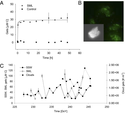
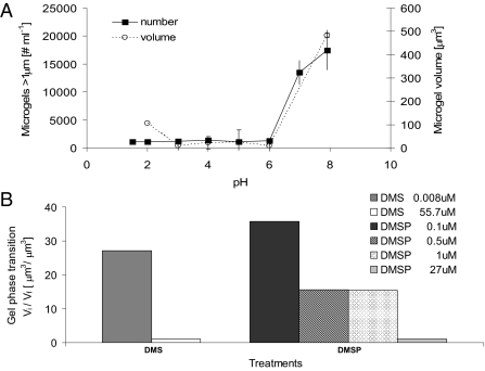
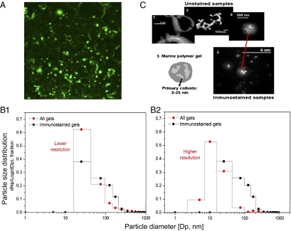
Marine microgels play an important role in regulating ocean basin-scale biogeochemical dynamics. In this paper, we demonstrate that, in the high Arctic, marine gels with unique physicochemical characteristics originate in the organic material produced by ice algae and/or phytoplankton in the surface water. The polymers in this dissolved organic pool assembled faster and with higher microgel yields than at other latitudes. The reversible phase transitions shown by these Arctic marine gels, as a function of pH, dimethylsulfide, and dimethylsulfoniopropionate concentrations, stimulate the gels to attain sizes below 1 μm in diameter. These marine gels were identified with an antibody probe specific toward material from the surface waters, sized, and quantified in airborne aerosol, fog, and cloud water, strongly suggesting that they dominate the available cloud condensation nuclei number population in the high Arctic (north of 80°N) during the summer season. Knowledge about emergent properties of marine gels provides important new insights into the processes controlling cloud formation and radiative forcing, and links the biology at the ocean surface with cloud properties and climate over the central Arctic Ocean and, probably, all oceans.


Conflict of interest statement

The authors declare no conflict of interest.

Figures

Fig. 1.
Fig. 1.
Polymer gel assembly as a function of time in high Arctic surface waters. (A) We monitored the assembly of polymer gels by measuring the percentage of polymers assembled as microgels at 4 °C (triangles). Control experiments in which Ca2+ was chelated from seawater with 10 mM EDTA showed no assembled gels, regardless of the time of observation (squares). Each point corresponds to the average of three replicates. We note that the assembly occurred very quickly, with the concentration and size of assembled polymer gels reaching equilibrium in 6 h. An average yield of assembly equal to 32% of the polymers in the DOM pool was measured for either SSW or SML water samples. (B) Microgels. Gels stained with chlortetracycline indicate the presence of bound Ca2+; insert at higher magnification. (C) Temporal variability in microgel concentration. During the drift phase of ASCOS at 87°N, we measured microgels in the SSW and SML in units of carbon, whereas we counted microgels in clouds with flow cytometry (as discrete particles) due to the large sample volume required to accurately measure carbon concentration in the microgels. SML microgels were enriched up to a factor of 5 with respect to SSW concentrations.
Fig. 2.
Fig. 2.
Microgel volume phase transition. (A) pH. Marine polymer gels can undergo a fast reversible volume phase transition (<1 min) from a swollen or hydrated phase to a condensed and collapsed phase by changing the pH of the sea water with H2SO4. We then stained the polymer gels with chlortetracycline, monitored the size of the polymer gels by confocal microscopy, and monitored the number of gels by flow cytometry. The swelling/condensation transition is reversible and has a steep sigmoidal change in the volume of the gels. Each data point corresponds to the average and SD of three samples. (B) DMS and DMSP. Marine polymer gels can undergo a fast, reversible change from a swelled/hydrated phase to a condensed phase as a function of DMS and DMSP concentrations, expressed as the ratio between initial (Vi) and final (Vf) microgel volume before and after adding the inducing compound, respectively, and measured with confocal microscopy.
Fig. 3.
Fig. 3.
Microgels in clouds. (A) Immunostained gels. Nanometer (nanogels)- and micrometer (microgels)-sized polymer gels immunostained with a specific antibody developed toward polymeric material collected in SML and SSW. (B) Gel size distribution from two independently analyzed subsamples. B1, Relative frequency of particle number (Np) [dNp/log(dDp): delta of Np/log delta particle diameter (Dp)] in a specific size range for unstained nanogels observed with FESEM (circles) and immunostained gels observed with confocal microscopy (squares), with instruments held at a similar, lower resolution. B2, The higher resolution of the FESEM allows the measurement of nanogels smaller than 10 nm. (C) Structure of nano and microgels. (C1) Low water-soluble organic particles in the SML present large quantities of colloidal-sized nanogels (<1 μm), which annealed into microgels bigger than 3 μm. (C2) Colloidal-sized nanogels tend to present fractal structures in sizes generally <200 nm and always smaller than 1 μm in both SML and cloud samples. (C3 and C4) The gels in the cloud samples present average sizes between 200 and 700 nm, with nanogels partitioned inside of both nonstained and immunostained particles. (C5) The schematic illustration shows that colloidal-sized nanogels tend to present a fractal structure, with a diffuse organic surface.

References

    1. Solomon S, et al. Climate change 2007: The physical science basis, contribution of working group 1 to the fourth assessment report of the Intergovernmental Panel on Climate Change. In: Solomon S, et al., editors. Intergovernmental Panel on Climate Change. New York, NY: Cambridge University Press; 2007. p. 996.
    1. Koåhler H. The nucleus in and the growth of hygroscopic droplets. Trans Faraday Soc. 1936;32:1152–1161.
    1. Intrieri J, et al. An annual cycle of Arctic surface cloud forcing at SHEBA. J Geophys Res Oceans. 2002;107 10.1029/2000JC000439.
    1. Leck C, Persson C. Seasonal and short-term variability in dimethyl sulfide, sulfur dioxide and biogenic sulfur and sea salt aerosol particles in the arctic marine boundary layer during summer and autumn. Tellus. 1996;48B:272–299.
    1. Leck C, Bigg K. Biogenic particles in the surface microlayer and overlaying atmosphere in the central Arctic Ocean during summer. Tellus. 2005;57B:305–316.

Publication types