. 2011 Aug 16;108(33):13456-61.
doi: 10.1073/pnas.1111218108. Epub 2011 Aug 8.

Phenotype-information-phenotype cycle for deconvolution of combinatorial antibody libraries selected against complex systems

Affiliations

Phenotype-information-phenotype cycle for deconvolution of combinatorial antibody libraries selected against complex systems

Hongkai Zhang et al. Proc Natl Acad Sci U S A. .

Abstract

Use of large combinatorial antibody libraries and next-generation sequencing of nucleic acids are two of the most powerful methods in modern molecular biology. The libraries are screened using the principles of evolutionary selection, albeit in real time, to enrich for members with a particular phenotype. This selective process necessarily results in the loss of information about less-fit molecules. On the other hand, sequencing of the library, by itself, gives information that is mostly unrelated to phenotype. If the two methods could be combined, the full potential of very large molecular libraries could be realized. Here we report the implementation of a phenotype-information-phenotype cycle that integrates information and gene recovery. After selection for phage-encoded antibodies that bind to targets expressed on the surface of Escherichia coli, the information content of the selected pool is obtained by pyrosequencing. Sequences that encode specific antibodies are identified by a bioinformatic analysis and recovered by a stringent affinity method that is uniquely suited for gene isolation from a highly degenerate collection of nucleic acids. This approach can be generalized for selection of antibodies against targets that are present as minor components of complex systems.


Conflict of interest statement

The authors declare no conflict of interest.

Figures

Fig. 1.
Fig. 1.
Phenotype-information-phenotype cycle. The cycle starts with selections from a combinatorial antibody library displayed on the surface of phage. The phage library is selected against two populations of bacteria that either display the antigen or serve as control cells (phage specific for antigen are in the minority and are colored red and other phage are indicated in blue). After selection, bound phage are eluted and their phagemids (circles) are analyzed by deep sequencing. The sequencing results from paired samples were compared by a bioinformatics analysis and DNA representing sequences overrepresented in phage that bind to the antigen presenting E. coli are extracted. A biotinylated probe whose design is based on the VH CDR3 of interest is synthesized and hybridized in solution with single-stranded circular DNA isolated from phage particles. The ssDNA selected by hybridization is captured on magnetic streptavidin beads and released by heating. The ssDNA is converted to dsDNA before transformation of suitable host E. coli cells.
Fig. 2.
Fig. 2.
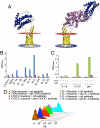
Antigen display on bacterial surfaces. Structural models (A, Left) The Lpp leader sequence and first nine amino acids of the E. coli major outer membrane lipoprotein (OmpA) was used to attach a variety of proteins to the E. coli surface. In this system, the posttranslational tripalmitoyl-S-glyceryl-cysteine component of the lipoprotein anchors the complex by inserting into the E. coli surface membrane. IL-6 was fused to the C terminus of OmpA (amino acids 46–159) and displayed on the surface of E. coli cells. (A, Right) Alternatively, IL-6 was fused to the C terminus of the MBP and the fusion product was anchored to the cell wall by linking it to the C terminus of OmpA. The view was generated using PyMOL. Protein Data Bank codes are 1ALU for IL-6, 3PGF for MBP, and 1QJP for OmpA. The IL-6 is represented as a blue ribbon diagram; MBP is rendered in magenta; and OmpA is colored yellow. The red and blue slabs represent the exoplasmic and cytoplasmic surface, respectively. (B) Expression of Lpp-OmpA-cytokine fusions. Cytokines were fused directly to the C terminus of OmpA. After induction at 22 °C for 3 h, the FLAG tag at the C terminus of fusion protein was detected by HRP-conjugated anti-FLAG antibody (blue) and compared to a control antibody of the same isotype (red). (C) Comparison of cytokine display level in the presence or absence of the MBP. IL-1β, chemokine (C-C motif) ligand 28 (CCL28), and gp41 antigens were displayed in duplicate, either fused directly to OmpA or fused to MBP–OmpA. The cytokines on the cell surface were detected by cell-ELISA with HRP conjugated to anti-FLAG antibody (blue and green) and compared to a control antibody of the same isotype (red and purple). (D) Detection of displayed cytokines by flow cytometry After induction, cells were stained with phycoerythrin (PE)-labeled cytokine specific antibodies for 1h, washed, and 10,000 E. coli events were analyzed with a Becton Dickinson flow cytometer. Each histogram is represented in pairs representing the specific versus control antibodies as indicated in the legend in the figure.
Fig. 3.
Fig. 3.
Selection of IL-1RA antibodies from a spiked-in library. Phage encoding the IL-1RA binding scFv (H9) were spiked into a ScFv naïve combinatorial antibody library containing 3.0 × 109 members at a ratio of one H9 encoding phage to 109 irrelevant phage. Two rounds of selection were carried out using a subtractive panning format in which at each round phage were first incubated with control bacteria and those that did not bind were next selected on bacteria displaying the IL-1RA antigen. The PCR-amplified VH repertoires from phage that bound to the paired samples at each round were sequenced using Roche 454 pyrosequencing. The VH CDR3s were identified and the frequency of each unique VH CDR3 was determined for the paired samples from the same round of selection (control versus experimental) to obtain a ratio of frequencies. In addition to the spiked-in H9 clone, we noted selective appearance of another clone (H1). (A) All CDR3s that had greater than 10 reads in at least one sample (ca. 0.3%) were ordered by similarity. The phylogenetic tree was constructed based on multiple sequence alignments via MAFFT and ClustalW2 tree generation methods (Left). The log2-fold change in frequency of the first round (blue) and the second round (red), and Z score of the first round (green) and the second round (purple) are shown (Right). Arrows indicate clones H1 and H9 (Right). (B) The sequences with highest differential ratio were recovered with overlap PCR and corresponding phages were prepared. The degree of binding to IL-1RA was tested by phage-ELISA using different concentrations of three different phage clones (H1, H9, and irrelevant). Phage were added to IL-1RA or BSA-coated wells. After washing, the bound phage were detected with HRP-conjugated phage antibody. (C) The scFvs were converted to full-length IgG that was produced in HEK293F cells and purified by protein G chromatography. Different concentrations of IgG were added to IL-1RA using BSA as a control protein. The bound antibody was detected with an HRP-conjugated antibody to the human IgG.
Fig. 4.
Fig. 4.
Epitope-directed antibody discovery. (A) Structure of IL-6. The structural representation was generated by PyMol. The Protein Data Bank code is 1ALU. The structure on the left represents IL-6 with amino acids 1–27 truncated (Δ 1–27). The cartoon on the right represents WT IL-6. The N-terminal 1–27 amino acids are highlighted in red and shown as ribbon diagrams. (B) Detection of wild-type and truncated IL-6 displayed on bacterial surfaces by flow cytometry. Wild-type and truncated IL-6 (Δ1–27) were displayed on the bacterial cell surface by fusing them to the C terminus of OmpA (Fig. 2). The induced bacteria were stained separately with two phycoerythrin (PE)-labeled mAbs. Monoclonal antibody 1 (R&D, catalog number IC206P) recognizes both the full-length and truncated version of IL-6, whereas mAb 2 (Biolegend, catalog number 501106) only recognizes the full-length version of the molecule. Irrelevant antibodies of the same isotype were used for control studies. After 1 h incubation at 24 °C, bacteria were washed and analyzed by flow cytometry. (C) Phylogenetic tree of CDR3 sequences from pyrosequencing. The PCR-amplified VH encoding DNA from phage bound to bacteria displaying WT or truncated IL-6 (Δ1–27) from the second round of selection was subjected to pyrosequencing. After a bioinformatics analysis, all CDR3 sequences observed greater than two times in at least one sample (ca. 0.1% frequency) were ordered by similarity. The phylogenetic tree on the left was constructed based on multiple sequence alignment via MAFFT and ClustalW2 tree generation methods. The log2-fold change in frequency (blue) and Z score (red) are shown on the right. (D) Binding of scFv to full-length and truncated IL-6 in phage-ELISA. Different concentrations of scFv displayed on phage were added to plates coated either with full-length or truncated human IL-6 (hIL-6) or BSA. The truncated IL-6 was prepared from HEK293F cells transfected with the truncated IL-6 overexpression vector and was purified on an anti-FLAG-tag column. After 1 h incubation at 37 °C, the wells were washed five times with PBS, after which HRP-conjugated antiphage antibody was added. Incubation was for 1 h at 37 °C. After five washes with PBS, 2,2′-azinobis(3-ethylbenzthiazoline-6-sulfonic acid) was added and the OD at 405 nm was measured. (E) Competitive ELISA to validate the epitope to which the selected antibody bound. ScFv protein N27-1 was purified from the bacterial culture supernatant and periplasmic space with a anti-FLAG antibody column. Increasing concentrations of the commercial IL-6 binding antibody (R&D, mAb2061), which we determined bound to the N terminus of the protein (Fig. 4B), were mixed with a constant concentration (1 μg/mL) of the selected scFv N27-1 and the mixtures were added to wells coated with recombinant hIL-6 (blue line). An irrelevant mAb against IL-1RA was used as control (red line). HRP-conjugated anti-FLAG antibody was added to detect the scFv. ScFv binding to BSA is shown as a green dashed line. The curves represent the concentration-dependent competitive inhibition of the signal generated by scFv binding by either the commercial mAb that bound to the N terminus of IL-6 (blue) or an irrelevant antibody (red).

References

    1. Edelman G, Gally JA. Degeneracy and complexity in biological systems. Proc Natl Acad Sci USA. 2001;98:13763–13768. - PMC - PubMed
    1. Huse WD, et al. Generation of a large combinatorial library of the immunoglobulin repertoire in phage lambda. Science. 1989;246:1275–1281. - PubMed
    1. Orlandi R, Güssow DH, Jones PT, Winter G. Cloning immunoglobulin variable domains for expression by te polymerase chain reaction. Proc Natl Acad Sci USA. 1989;86:3833–3837. - PMC - PubMed
    1. Sastry L, et al. Cloning of the immunological repertoire in Escherichia coli for generation of monoclonal catalytic antibodies: Construction of a heavy chain variable region-specific cDNA library. Proc Natl Acad Sci USA. 1989;86:5728–5732. - PMC - PubMed
    1. McCafferty J, Griffiths AD, Winter G, Chiswell DJ. Phage antibodies: Filamentous phage displaying antibody variable domains. Nature. 1990;348:552–554. - PubMed

Publication types

MeSH terms

LinkOut - more resources