. 2011 Aug 23;108(34):14103-8.
doi: 10.1073/pnas.1108799108. Epub 2011 Aug 8.

Iduna is a poly(ADP-ribose) (PAR)-dependent E3 ubiquitin ligase that regulates DNA damage

Affiliations

Iduna is a poly(ADP-ribose) (PAR)-dependent E3 ubiquitin ligase that regulates DNA damage

Ho Chul Kang et al. Proc Natl Acad Sci U S A. .

Abstract

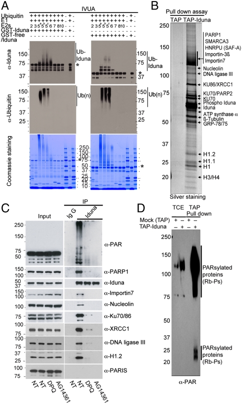
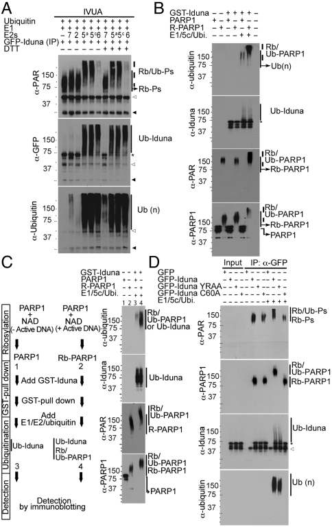
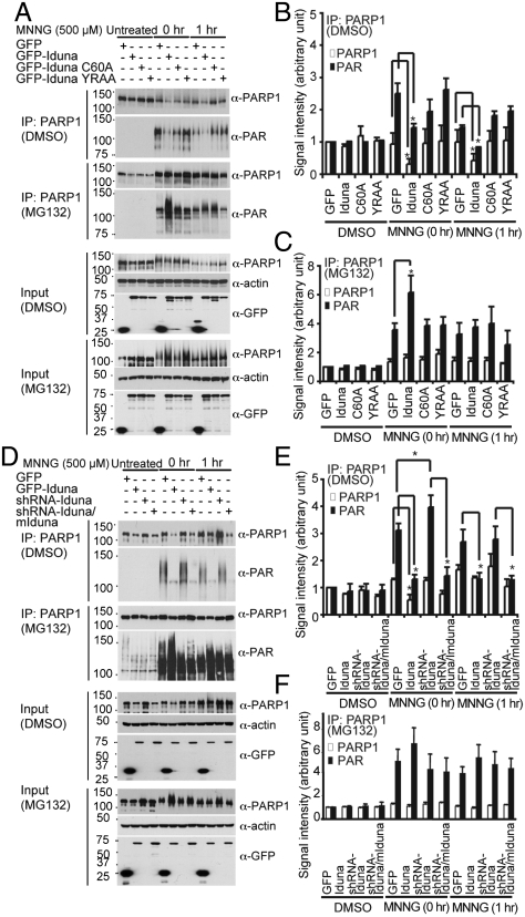
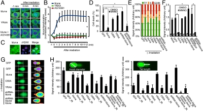
Ubiquitin mediated protein degradation is crucial for regulation of cell signaling and protein quality control. Poly(ADP-ribose) (PAR) is a cell-signaling molecule that mediates changes in protein function through binding at PAR binding sites. Here we characterize the PAR binding protein, Iduna, and show that it is a PAR-dependent ubiquitin E3 ligase. Iduna's E3 ligase activity requires PAR binding because point mutations at Y156A and R157A eliminate Iduna's PAR binding and Iduna's E3 ligase activity. Iduna's E3 ligase activity also requires an intact really interesting new gene (RING) domain because Iduna possessing point mutations at either H54A or C60A is devoid of ubiquitination activity. Tandem affinity purification reveals that Iduna binds to a number of proteins that are either PARsylated or bind PAR including PAR polymerase-1, 2 (PARP1, 2), nucleolin, DNA ligase III, KU70, KU86, XRCC1, and histones. PAR binding to Iduna activates its E3 ligase function, and PAR binding is required for Iduna ubiquitination of PARP1, XRCC1, DNA ligase III, and KU70. Iduna's PAR-dependent ubiquitination of PARP1 targets it for proteasomal degradation. Via PAR binding and ubiquitin E3 ligase activity, Iduna protects against cell death induced by the DNA damaging agent N-methyl-N-nitro-N-nitrosoguanidine (MNNG) and rescues cells from G1 arrest and promotes cell survival after γ-irradiation. Moreover, Iduna facilitates DNA repair by reducing apurinic/apyrimidinic (AP) sites after MNNG exposure and facilitates DNA repair following γ-irradiation as assessed by the comet assay. These results define Iduna as a PAR-dependent E3 ligase that regulates cell survival and DNA repair.


Conflict of interest statement

The authors declare no conflict of interest.

Figures

Fig. 1.
Fig. 1.
Iduna is an ubiquitin E3 ligase that binds PARsylated proteins. (A) Screening of UbcH E2 enzymes for Iduna via an in vitro ubiquitination assay (IVUA) with GST-Iduna (Left) or GST free Iduna (Right). Samples were resolved in 8–16% SDS/PAGE and either stained with coomassie or immunoblotted by anti-Iduna or anti-ubiquitin antibody. (*) indicates unmodified GST-Iduna or GST-free Iduna. (B) Identification of potential Iduna substrates. TAP purification of SK-N-SH cells stably transfected with pNTAP or pNATP-Iduna were resolved in 8–16% SDS/PAGE and silver stained. Mass spectrometric analysis identified 16 proteins as indicated. (C) Iduna interacts with its potential substrates in a PAR-dependent manner. MCF7 cells were preincubated with DMSO or PARP1 inhibitors as indicated and then harvested and lysed. Endogenous Iduna was immunoprecipitated by anti-Iduna antibody from each cell lysate and subjected into immunoblot with appropriate antibodies. IgG was used as a negative control. (D) Iduna strongly binds to PARsylated proteins. TAP or TAP-Iduna pull-down samples were analyzed by immunoblot with anti-PAR antibody. Abbreviations: Ub (n), polyubiquitin chains; Ub-Iduna, poly ubiquitinated Iduna. All experiments were repeated two to three times.
Fig. 2.
Fig. 2.
Iduna mediates PARsylation-dependent ubiquitination of its substrates. (A) In vitro ubquitination assay of immunoprecipiated GFP-Iduna and different UbcHE2 enzymes in presence or absence of DTT as indicated. Samples were analyzed by immunoblot with anti-PAR, anti-GFP, and anti-ubiquitin antibodies. White or black arrow heads indicate the immunoglobulin heavy or light chains, respectively. (B) In vitro ubiquitination assay of recombinant PARP1 or PARsylated PARP1 (R-PARP1) by GST-Iduna subjected to immunoblot analysis with indicated antibodies. (C) Iduna binds and/or ubiquitinates PARP1 in a PARsylation-dependent manner. PARP1 or R-PARP1 were incubated with GST-Iduna, followed by GST pull-down and subjected to the in vitro ubiquitination assay (Left) and analyzed by immunoblot (Right). (D) Iduna is a PAR-dependent ubiquitin E3 ligase. In vitro ubiquitination assay of immnuoprecipitated GFP, GFP-Iduna, GFP-Iduna YRAA, and GFP-Iduna C60A analyzed by immunoblot with anti-GFP, anti-ubiquitin, anti-PAR, anti-PARP1, and anti-ubiquitin antibodies. Abbreviations: Rb-P, PARsylated proteins; Rb/Ub-P, PARsylated and polyubiquitinated proteins; Ub (n), polyubiquitin chains; Rb/Ub-PARP1, PARsylated and polyubiquitinated PARP1; Ub-Iduna, poly ubiquitinated Iduna; Rb-PARP1, PARsylated PARP1. All experiments were repeated three times.
Fig. 3.
Fig. 3.
PARsylation-dependent PARP1 degradation by Iduna. (A) Stable MCF7 cell lines expressing GFP, GFP-Iduna, GFP-Iduna C60A, or GFP-Iduna YRAA were exposed to DMSO or MNNG (500 μM) for 15 min with or without MG132. PARP1 was immunoprecipitated at 0 or 1 hr after the MNNG challenge. PARP1 and PARsylated-PARP1 were monitored by immunoblot with anti-PARP1 and anti-PAR antibodies. (B) Quantification of PARP1 and PARsylated-PARP1 in the absence of MG132. (C) Quantification of PARP1 and PARsylated-PARP1 in presence of MG132. Quantifications were normalized with respect to actin levels. (D) Levels of immunoprecipitated PARP1 and PARsylated PARP1 after exposure to DMSO or MNNG (500 μM) for 15 min with or without MG132 GFP in MCF7 cell lines stably expressing GFP-Iduna, shRNA-Iduna, or shRNA-Iduna/GFP-mouse Iduna (mIduna) at 0 or 1 hr after the MNNG challenge. (E) Quantification of PARP1 and PARsylated PARP1 normalized to actin in absence of MG132. (F) Quantification of the PARP1 and PARsylated-PARP1 normalized to actin in presence of MG132. Data represents mean ± s.e.m., n = 3, * P < 0.05 by ANOVA with Tukey-Kramer’s post hoc test. All experiments were repeated two to three times.
Fig. 4.
Fig. 4.
Iduna protects against DNA damage. (A) Recruitment of stably expressed GFP-Iduna to sites of laser (405 nm) microirradiation induced DNA damage in MCF7 cells. GFP-Iduna YRAA does not translocate to the damage site. The PARP inhibitor AG14361 blocks GFP-Iduna recruitment. (B) Comparative quantitative analysis of GFP-Iduna, GFP-Iduna-YRAA, and GFP-Iduna plus PARP inhibitor AG14361 kinetics after DNA damage. (C) GFP-Iduna localizes to sites of DNA damage as indicated by colocalization with γH2AX immunostaining. (D) Stable MCF7 cell lines expressing GFP, GFP-Iduna, GFP-Iduna C60A, GFP-Iduna YRAA , shRNA-Iduna, or shRNA-Iduna/GFP-mouse Iduna (mIduna) were treated with DMSO or MNNG (500 μM) for 15 min. After 24 hr, the cells were stained with Hoechst 33342 and propidium iodide (PI), and dead cells were counted by automated computer-assisted program. (E) Stable MCF7 cell lines were γ-irradiated at 2 Gy as indicated. Cells were collected 16 hr after irradiation and then DNA content was measured by flow cytometry. The percentage of each cell cycle phase was measured by FlowJo software using the Dean-Jett-Fox model. (F) Stable MCF7 lines as indicated were treated with either DMSO or MNNG. After 1 h, genomic DNA was isolated and then AP sites on genomic DNA were labeled with biotin by Aldehyde Reactive Probe (ARP) reagent. Biotin-labeled AP sites were quantified using an avidin—biotin assay. (G) Stable MCF7 lines were γ-irradiated at 2 Gy as indicated. After 15 min, cells were collected and then subjected to the comet assay. (H) Quantification of head diameter after comet assay. (I) Quantification of tail length after comet assay. Data represents mean ± s.e.m., n = 3, * P < 0.05 by ANOVA with Tukey-Kramer’s post hoc test. All experiments were repeated three to four times.

References

    1. Ciechanover A. The ubiquitin-proteasome pathway: On protein death and cell life. EMBO J. 1998;17:7151–7160. - PMC - PubMed
    1. Di Fiore PP, Polo S, Hofmann K. When ubiquitin meets ubiquitin receptors: A signalling connection. Nat Rev. 2003;4:491–497. - PubMed
    1. Hochstrasser M. New structural clues to substrate specificity in the “ubiquitin system”. Mol Cell. 2002;9:453–454. - PubMed
    1. Harper JW. A phosphorylation-driven ubiquitination switch for cell-cycle control. Trends Cell Biol. 2002;12:104–107. - PubMed
    1. Prudden J, et al. SUMO-targeted ubiquitin ligases in genome stability. EMBO J. 2007;26:4089–4101. - PMC - PubMed

Publication types

MeSH terms

Substances