. 2012 Feb 21;109(8):2796-801.
doi: 10.1073/pnas.1104303108. Epub 2011 Aug 8.

Leukocyte composition of human breast cancer

Affiliations

Leukocyte composition of human breast cancer

Brian Ruffell et al. Proc Natl Acad Sci U S A. .

Abstract

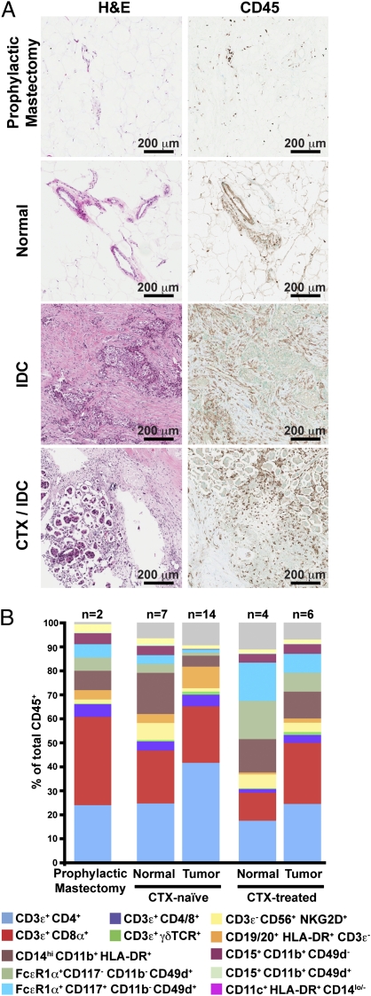
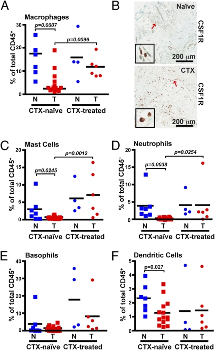
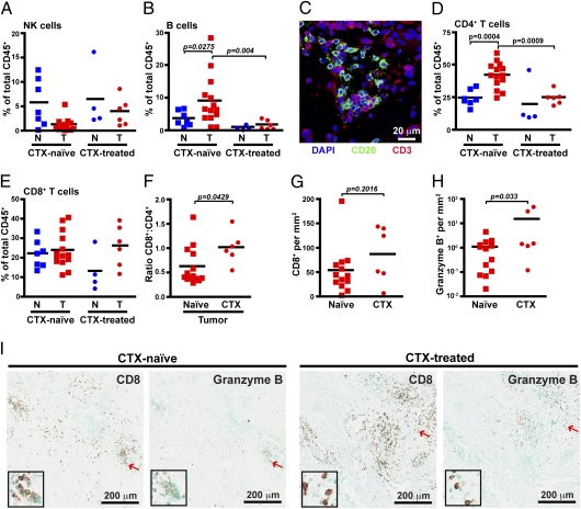
Retrospective clinical studies have used immune-based biomarkers, alone or in combination, to predict survival outcomes for women with breast cancer (BC); however, the limitations inherent to immunohistochemical analyses prevent comprehensive descriptions of leukocytic infiltrates, as well as evaluation of the functional state of leukocytes in BC stroma. To more fully evaluate this complexity, and to gain insight into immune responses after chemotherapy (CTX), we prospectively evaluated tumor and nonadjacent normal breast tissue from women with BC, who either had or had not received neoadjuvant CTX before surgery. Tissues were evaluated by polychromatic flow cytometry in combination with confocal immunofluorescence and immunohistochemical analysis of tissue sections. These studies revealed that activated T lymphocytes predominate in tumor tissue, whereas myeloid lineage cells are more prominant in "normal" breast tissue. Notably, residual tumors from an unselected group of BC patients treated with neoadjuvant CTX contained increased percentages of infiltrating myeloid cells, accompanied by an increased CD8/CD4 T-cell ratio and higher numbers of granzyme B-expressing cells, compared with tumors removed from patients treated primarily by surgery alone. These data provide an initial evaluation of differences in the immune microenvironment of BC compared with nonadjacent normal tissue and reveal the degree to which CTX may alter the complexity and presence of selective subsets of immune cells in tumors previously treated in the neoadjuvant setting.


Conflict of interest statement

The authors declare no conflict of interest.

Figures

Fig. 1.
Fig. 1.
Leukocyte infiltration of human breast tumors. (A) Hematoxylin and eosin (H&E) staining of tissue sections (Left) with representative immunohistochemistry for CD45 (Right) shown for each. (B) Flow cytometric analysis of leukocyte populations within human breast tumors. Results are shown as a percent of total CD45+ cells with markers used to define specific lineages shown below.
Fig. 2.
Fig. 2.
Increased myeloid-lineage leukocyte infiltration within CTX-treated patients. (A) CD14hiCD11b+HLA-DR+ macrophages shown as a percent of total CD45+ cells as determined by flow cytometry. (B) Representative immunohistochemistry for CSF1R in tumors from either CTX-naïve (Upper) or CTX-treated (Lower) patients. Red arrows indicate cells displayed in enlarged insets. FcεR1α+CD117+CD11bCD49d+ mast cells (C), CD15+CD11b+CD49d neutrophils (D), FcεR1α+CD117CD11bCD49d+ basophils (E), and CD11c+HLA-DR+CD14lo/− (F) DCs shown as a percent of total CD45+ cells. N, nonadjacent normal; T, tumor.
Fig. 3.
Fig. 3.
CD68 is not a specific macrophage marker in human breast tumor tissue. (A) Representative immunohistochemistry within tumors for CD68 (Left), CSF1R (Center), and CD163 (Right) in serial sections from a CTX-treated patient. Red arrows indicate cells displayed in enlarged insets. (B) Immunofluorescent staining of human breast tumors for CD68 (red) in conjunction with CSF1R (i and ii) or CD45 (iii and iv). (C) Immunofluorescent staining for CD68 (red) in conjunction with pan-keratin (green; i), or CD31 (green) and smooth muscle actin-α (SMA; purple; ii).
Fig. 4.
Fig. 4.
Tissue-infiltrating T cells display an activated phenotype. (A and B) Representative histograms of CD3+CD4+ (Upper) or CD3+CD8+ (Lower) T cells isolated from a single CTX-treated patient with both normal (blue) and tumor (red) tissue. Expression of activation markers CD69 (Left), HLA-DR (Center Left), CD45RA (Center Right), and CD27 (Right) are shown in A, and expression of chemokine receptors CCR4 (Left), CCR5 (Center Left), CCR7 (Center Right) and CXCR3 (Right) are shown in B.
Fig. 5.
Fig. 5.
Improved cytotoxic T-cell response in CTX-treated tumors. CD3ε CD56+NKG2D+ natural killer cells (A) and CD3εCD19/20+HLA-DR+ B cells (B) shown as a percent of total CD45+ cells as determined by flow cytometry. (C) Immunofluorescent staining of tumors for CD20 (green) and CD3 (red). CD3ε+CD4+ T cells (D) and CD3ε+CD8+ T cells (E) are shown as a percent of total CD45+ cells. (F) Ratio of CD8+ to CD4+ T cells within CTX-naive versus CTX-treated tumors. Number of CD8-positive (G) and granzyme B-positive (H) cells per area as determined by automated counting. (I) Representative sections stained with CD8 or granzyme B from CTX-naive (Left) or CTX-treated (Right) tumors. Red arrows indicate cells displayed in enlarged insets. N, nonadjacent normal; T, tumor.

References

    1. DeNardo DG, et al. CD4(+) T cells regulate pulmonary metastasis of mammary carcinomas by enhancing protumor properties of macrophages. Cancer Cell. 2009;16:91–102. - PMC - PubMed
    1. Ménard S, et al. Lymphoid infiltration as a prognostic variable for early-onset breast carcinomas. Clin Cancer Res. 1997;3:817–819. - PubMed
    1. Pupa SM, et al. Macrophage infiltrate and prognosis in c-erbB-2-overexpressing breast carcinomas. J Clin Oncol. 1996;14:85–94. - PubMed
    1. Bingle L, Brown NJ, Lewis CE. The role of tumour-associated macrophages in tumour progression: Implications for new anticancer therapies. J Pathol. 2002;196:254–265. - PubMed
    1. Mukhtar RA, Nseyo O, Campbell MJ, Esserman LJ. Tumor-associated macrophages in breast cancer as potential biomarkers for new treatments and diagnostics. Expert Rev Mol Diagn. 2011;11:91–100. - PubMed

Publication types

MeSH terms