

A framework of vertebra segmentation using the active shape model-based approach
- PMID: 21826134
- PMCID: PMC3149802
- DOI: 10.1155/2011/621905
A framework of vertebra segmentation using the active shape model-based approach
Abstract
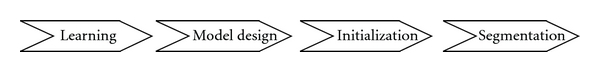
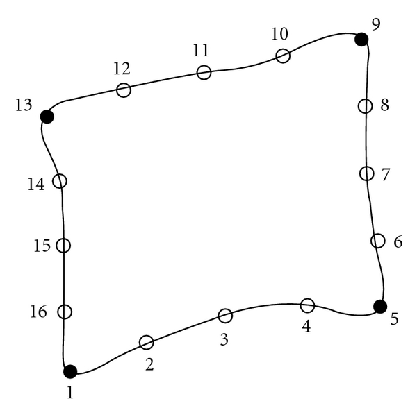
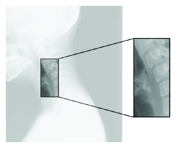
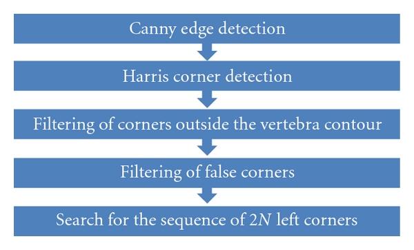
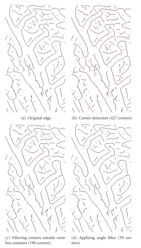
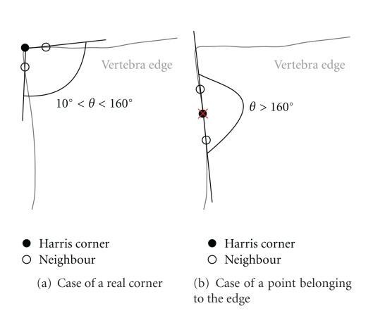
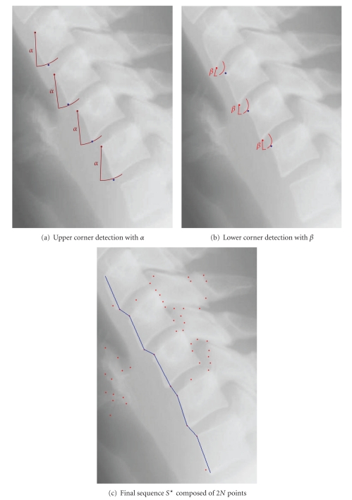
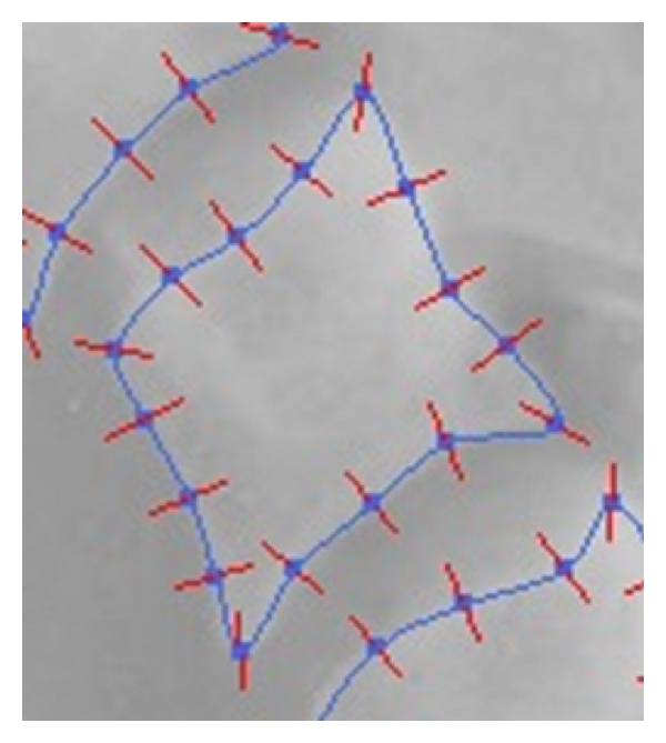
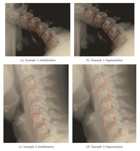
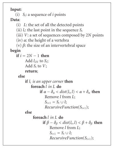
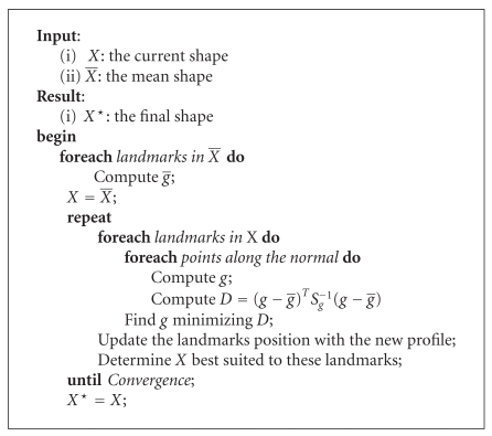
We propose a medical image segmentation approach based on the Active Shape Model theory. We apply this method for cervical vertebra detection. The main advantage of this approach is the application of a statistical model created after a training stage. Thus, the knowledge and interaction of the domain expert intervene in this approach. Our application allows the use of two different models, that is, a global one (with several vertebrae) and a local one (with a single vertebra). Two modes of segmentation are also proposed: manual and semiautomatic. For the manual mode, only two points are selected by the user on a given image. The first point needs to be close to the lower anterior corner of the last vertebra and the second near the upper anterior corner of the first vertebra. These two points are required to initialize the segmentation process. We propose to use the Harris corner detector combined with three successive filters to carry out the semiautomatic process. The results obtained on a large set of X-ray images are very promising.
Figures


Similar articles
-
Spine localization in X-ray images using interest point detection.J Digit Imaging. 2009 Jun;22(3):309-18. doi: 10.1007/s10278-007-9099-3. Epub 2008 Feb 14. J Digit Imaging. 2009. PMID: 18273669 Free PMC article.
-
VolHOG: a volumetric object recognition approach based on bivariate histograms of oriented gradients for vertebra detection in cervical spine MRI.Med Phys. 2014 Aug;41(8):082305. doi: 10.1118/1.4890587. Med Phys. 2014. PMID: 25086554
-
A semiautomatic segmentation method for prostate in CT images using local texture classification and statistical shape modeling.Med Phys. 2018 Jun;45(6):2527-2541. doi: 10.1002/mp.12898. Epub 2018 Apr 23. Med Phys. 2018. PMID: 29611216 Free PMC article.
-
Fully automatic cervical vertebrae segmentation framework for X-ray images.Comput Methods Programs Biomed. 2018 Apr;157:95-111. doi: 10.1016/j.cmpb.2018.01.006. Epub 2018 Jan 12. Comput Methods Programs Biomed. 2018. PMID: 29477438
-
Semiautomatic segmentation of vertebrae in lateral x-rays using a conditional shape model.Acad Radiol. 2007 Oct;14(10):1156-65. doi: 10.1016/j.acra.2007.06.003. Acad Radiol. 2007. PMID: 17889333 Review.
Cited by
-
An Automated Deep Learning Approach for Spine Segmentation and Vertebrae Recognition Using Computed Tomography Images.Diagnostics (Basel). 2023 Aug 12;13(16):2658. doi: 10.3390/diagnostics13162658. Diagnostics (Basel). 2023. PMID: 37627917 Free PMC article.
-
Cobb Angle Measurement of Spine from X-Ray Images Using Convolutional Neural Network.Comput Math Methods Med. 2019 Feb 19;2019:6357171. doi: 10.1155/2019/6357171. eCollection 2019. Comput Math Methods Med. 2019. PMID: 30996731 Free PMC article.
-
Self-learning computers for surgical planning and prediction of postoperative alignment.Eur Spine J. 2018 Feb;27(Suppl 1):123-128. doi: 10.1007/s00586-018-5497-0. Epub 2018 Feb 9. Eur Spine J. 2018. PMID: 29427011 Review.
-
A Region-Based Deep Level Set Formulation for Vertebral Bone Segmentation of Osteoporotic Fractures.J Digit Imaging. 2020 Feb;33(1):191-203. doi: 10.1007/s10278-019-00216-0. J Digit Imaging. 2020. PMID: 31011954 Free PMC article.
-
Automated 3D closed surface segmentation: application to vertebral body segmentation in CT images.Int J Comput Assist Radiol Surg. 2016 May;11(5):789-801. doi: 10.1007/s11548-015-1320-0. Epub 2015 Nov 11. Int J Comput Assist Radiol Surg. 2016. PMID: 26558791
References
-
- McInerney T, Terzopoulos D. Deformable models in medical image analysis: a survey. Medical Image Analysis. 1996;1(2):91–108. - PubMed
-
- Kass MA, Witkin A, Terzopoulos D. Snakes: active contour models. International Journal of Computer Vision. 1988;1(4):321–331.
-
- Xu C, Prince JL. Snakes, shapes, and gradient vector flow. IEEE Transactions on Image Processing. 1998;7(3):359–369. - PubMed
-
- McInerney T, Terzopoulos D. T-snakes: topology adaptive snakes. Medical Image Analysis. 2000;4(2):73–91. - PubMed
-
- Lobregt S, Viergever MA. Discrete dynamic contour model. IEEE Transactions on Medical Imaging. 1995;14(1):12–24. - PubMed
LinkOut - more resources
Full Text Sources
