Traumatic and Non-traumatic Osteonecrosis in the Femoral Head of a Rabbit Model
- PMID: 21826172
- PMCID: PMC3146007
- DOI: 10.5625/lar.2011.27.2.127
Traumatic and Non-traumatic Osteonecrosis in the Femoral Head of a Rabbit Model
Abstract
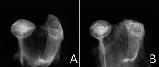
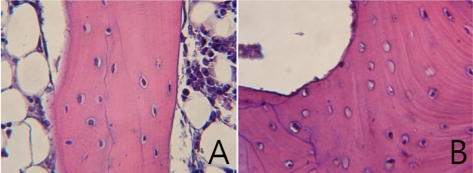
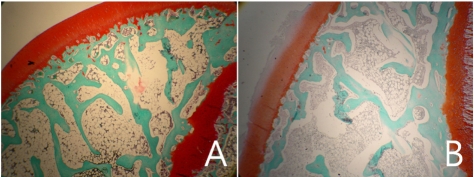
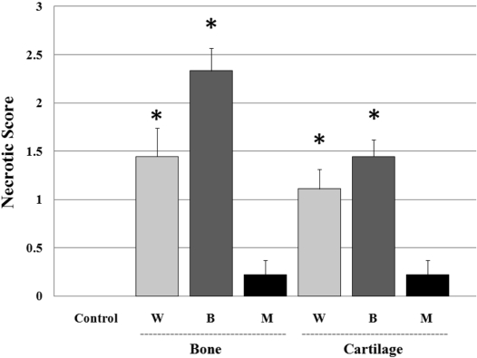
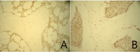
Osteonecrosis of the femoral head is an idiopathic, debilitating and progressive disease. A number of traumatic or non-traumatic animal models have been reported for research on osteonecrosis. This study was performed to compare the efficacy of femoral head osteonecrosis in rabbits by traumatic and non-traumatic methods. Twenty-seven New Zealand White rabbits were divided into three experimental groups, nine heads each. Two groups were surgically induced into osteonecrosis; a steel cerclage wire was ligated tightly around the neck of the right femoral head (Group W), and the femoral neck was tied with a cerclage wire in the same way as in the W group, and burned by attachment of an electrode tip to the wire and then the wire was removed (Group B). The other group was induced into osteonecrosis with a single intra-muscular injection of 20 mg/kg methyl-prednisolone acetate single injection (Group M). In the control group, the left femoral head of animals in group W and B was used. After two weeks, rabbits were sacrificed and the femoral head and neck were collected. Osteonecrosis of the femoral head was evaluated by radiography, histology and immunohistology methods. Osteonecrosis lesions in the femoral head were identified in traumatic models of groups W and B. Cartilage degeneration in the superficial layer and TUNEL positive cells in the femoral head were detected more in Group B than in Group W. These findings revealed that short-term induced osteonecrosis of the femoral head was effectively achieved by cautery around the femoral neck.
Keywords: animal model; bone; cartilage; osteonecrosis; rabbit.
Figures
References
-
- Mont MA, Hungerford DS. Non-traumatic avascular necrosis of the femoral head. J Bone Joint Surg Am. 1995;77:459–474. - PubMed
-
- Trueta J. The normal vascular anatomy of the human femoral head during growth. J Bone Joint Surg Br. 1957;39-B:358–394. - PubMed
-
- Rokkanen P, Slatis P. Effect of compression on the healing of subcapital osteotomies of the femoral neck and on the avascularized femoral head. An experimental study on adult rabbits. Acta Orthop Scand. 1967;38:163–173. - PubMed
-
- Kenzora JE, Steele RE, Yosipovitch ZH. Experimental osteonecrosis of the femoral head in adult rabbits. Clin Orthop. 1978;130:8–46. - PubMed
-
- Rösingh GE, James J. Early phases of avascular necrosis of the femoral head in rabbits. J Bone Joint Surg Br. 1969;51:165–174. - PubMed