. 2011 Aug 9:12:32.
doi: 10.1186/1471-2121-12-32.

Hypoxia-mimetic agents inhibit proliferation and alter the morphology of human umbilical cord-derived mesenchymal stem cells

Affiliations

Hypoxia-mimetic agents inhibit proliferation and alter the morphology of human umbilical cord-derived mesenchymal stem cells

Hui-Lan Zeng et al. BMC Cell Biol. .

Abstract

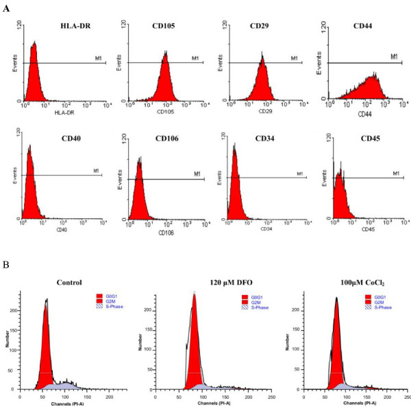
Background: The therapeutic efficacy of human mesenchymal stem cells (hMSCs) for the treatment of hypoxic-ischemic diseases is closely related to level of hypoxia in the damaged tissues. To elucidate the potential therapeutic applications and limitations of hMSCs derived from human umbilical cords, the effects of hypoxia on the morphology and proliferation of hMSCs were analyzed.

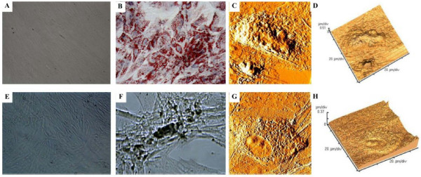
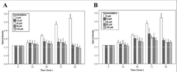
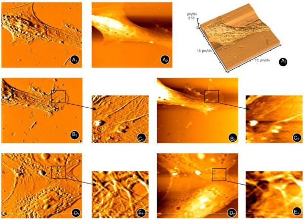
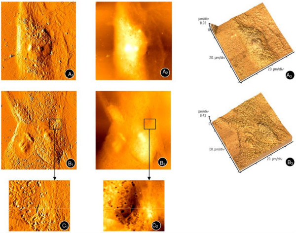
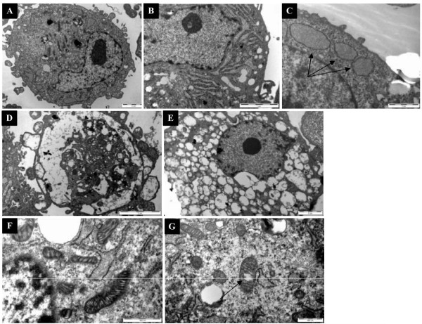
Results: After treatment with DFO and CoCl₂, hMSCs were elongated, and adjacent cells were no longer in close contact. In addition, vacuole-like structures were observed within the cytoplasm; the rough endoplasmic reticulum expanded, and expanded ridges were observed in mitochondria. In addition, DFO and CoCl₂ treatments for 48 h significantly inhibited hMSCs proliferation in a concentration-dependent manner (P < 0.05). This treatment also increased the number of cells in G0/G1 phase and decreased those in G2/S/M phase.

Conclusions: The hypoxia-mimetic agents, DFO and CoCl₂, alter umbilical cord-derived hMSCs morphology and inhibit their proliferation through influencing the cell cycle.


Figures

Figure 1
Figure 1
Morphological changes in hMSCs primary cultures over time. A) First passage at 5 days (100×). B) Third passage at 3 days (50×).
Figure 2
Figure 2
Histograms representing the immunophenotype and cell cycle distribution as determined by flow cytometry. The phenotype of hMSCs was determined after cell isolation (A). The cell cycle distribution was determined without (control) or with 120 μM DFO or with 100 μM CoCl2 for 48 h (B).
Figure 3
Figure 3
Adipogenic and osteogenic differentiation of hMSCs. Adipogenesis was detected by the formation of intracytoplasmic lipid droplets stained with oil red O (A, no induction; B, induced cells; 100×). Osteogenic differentiation was demonstrated by calcium deposition as evidenced von Kossa staining (E, no induction; F, induced cells; 100×). Atomic force microscopy of umbilical cord-derived hMSCs upon adipogenic (C, D) and osteogenic (G, H) differentiation after 21 days in normoxic conditions were shown as error signal images (C,G) and 3-dimension graphs (D,H).
Figure 4
Figure 4
Reduced hMSCs growth after treatment with hypoxia mimetics. A) hMSCs were treated with the indicated concentration of A) DFO or B) CoCl2 at various time points. Data represent the mean ± SD. *P < 0.05 as compared to the control group.
Figure 5
Figure 5
Atomic force microscopy of hMSCs under normoxic conditions. A typical long spindle, cytoskeleton (A1, A2, A3), and palpus-like or cicada wing-like pseudopodium (B1,B2, enlarged areas shown in C1 and C2 respectively) were observed. The mesh-like cytoskeleton of adjacent hMSCs (D1, D2) can also been seen (enlarged areas shown in E1, E2).
Figure 6
Figure 6
Atomic force microscopy of hMSCs upon treatment with hypoxia mimetics. hMSCs were treated with 120 μM DFO (A1, A2, A3) and 100 μM CoCl2 (B1, B2, B3) for 4 days. Enlarged areas of B1 and B2 are shown in C1 and C2 respectively.
Figure 7
Figure 7
Transmission electron microscopy of hMSCs. A) Complete view of untreated (8900×) or D and E) DFO-treated hMSCs after 3 days (8900×). (B and C) The endoplasmic reticulum of untreated hMSCs (24000×). F) The mitochondria of untreated (30000×) and G) DFO-treated hMSCs (30000×).

Similar articles

Cited by

References

    1. Friedenstein AJ, Chailakhyan RK, Latsinik NV, Panasyuk AF, Keiliss-Borok IV. Stromal cells responsible for transferring the microenvironment of the hemopoietic tissues, Cloning in vitro and retransplantation in vivo. Transplantation. 1974;17:331–40. doi: 10.1097/00007890-197404000-00001. - DOI - PubMed
    1. Majore I, Moretti P, Hass R, Kasper C. Identification of subpopulations in mesenchymal stem cell-like cultures from human umbilical cord. Cell Communication and Signaling. 2009;7:1–8. doi: 10.1186/1478-811X-7-1. - DOI - PMC - PubMed
    1. Alviano F, Fossati V, Marchionni C, Arpinati M, Bonsi L, Franchina M, Lanzoni G, Cantoni S, Cavallini C, Bianchi F, Tazzari PL, Pasquinelli G, Foroni L, Ventura C, Grossi A, Bagnara GP. Term Amniotic membrane is a high throughput source for multipotent Mesenchymal Stem Cells with the ability to differentiate into endothelial cells in vitro. BMC Dev Biol. 2007;7:1–14. doi: 10.1186/1471-213X-7-1. - DOI - PMC - PubMed
    1. Fukuchi Y, Nakajima H, Sugiyama D, Hirose I, Kitamura T, Tsuji K. Human placenta-derived cells have mesenchymal stem/progenitor cell potential. Stem Cells. 2004;22:649–58. doi: 10.1634/stemcells.22-5-649. - DOI - PubMed
    1. Secco M, Zucconi E, Vieira NM, Fogaça LL, Cerqueira A, Carvalho MD, Jazedje T, Okamoto OK, Muotri AR, Zatz M. Multipotent Stem Cells from Umbilical Cord: Cord Is Richer than Blood. Stem Cells. 2008;26:146–50. doi: 10.1634/stemcells.2007-0381. - DOI - PubMed

Publication types

LinkOut - more resources