

In vitro activation and enzyme kinetic analysis of recombinant midgut serine proteases from the Dengue vector mosquito Aedes aegypti
- PMID: 21827688
- PMCID: PMC3162888
- DOI: 10.1186/1471-2091-12-43
In vitro activation and enzyme kinetic analysis of recombinant midgut serine proteases from the Dengue vector mosquito Aedes aegypti
Abstract
Background: The major Dengue virus vector Aedes aegypti requires nutrients obtained from blood meal proteins to complete the gonotrophic cycle. Although bioinformatic analyses of Ae. aegypti midgut serine proteases have provided evolutionary insights, very little is known about the biochemical activity of these digestive enzymes.
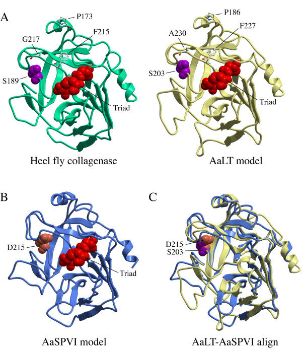
Results: We used peptide specific antibodies to show that midgut serine proteases are expressed as zymogen precursors, which are cleaved to the mature form after blood feeding. Since midgut protein levels are insufficient to purify active proteases directly from blood fed mosquitoes, we engineered recombinant proteins encoding a heterologous enterokinase cleavage site to permit generation of the bona fide mature form of four midgut serine proteases (AaET, AaLT, AaSPVI, AaSPVII) for enzyme kinetic analysis. Cleavage of the chromogenic trypsin substrate BApNA showed that AaET has a catalytic efficiency (k(cat)/K(M)) that is ~30 times higher than bovine trypsin, and ~2-3 times higher than AaSPVI and AaSPVII, however, AaLT does not cleave BApNA. To measure the enzyme activities of the mosquito midgut proteases using natural substrates, we developed a quantitative cleavage assay based on cleavage of albumin and hemoglobin proteins. These studies revealed that the recombinant AaLT enzyme was indeed catalytically active, and cleaved albumin and hemoglobin with equivalent efficiency to that of AaET, AaSPVI, and AaSPVII. Structural modeling of the AaLT and AaSPVI mature forms indicated that AaLT is most similar to serine collagenases, whereas AaSPVI appears to be a classic trypsin.
Conclusions: These data show that in vitro activation of recombinant serine proteases containing a heterologous enterokinase cleavage site can be used to investigate enzyme kinetics and substrate cleavage properties of biologically important mosquito proteases.
© 2011 Rascón et al; licensee BioMed Central Ltd.
Figures


Similar articles
-
Molecular genetic analysis of midgut serine proteases in Aedes aegypti mosquitoes.Insect Biochem Mol Biol. 2009 Dec;39(12):903-12. doi: 10.1016/j.ibmb.2009.10.008. Epub 2009 Nov 3. Insect Biochem Mol Biol. 2009. PMID: 19883761 Free PMC article.
-
Soluble expression of recombinant midgut zymogen (native propeptide) proteases from the Aedes aegypti Mosquito Utilizing E. coli as a host.BMC Biochem. 2018 Dec 18;19(1):12. doi: 10.1186/s12858-018-0101-0. BMC Biochem. 2018. PMID: 30563449 Free PMC article.
-
Expression profiling and comparative analyses of seven midgut serine proteases from the yellow fever mosquito, Aedes aegypti.J Insect Physiol. 2010 Jul;56(7):736-44. doi: 10.1016/j.jinsphys.2010.01.003. Epub 2010 Feb 2. J Insect Physiol. 2010. PMID: 20100490 Free PMC article.
-
Comprehensive proteolytic profiling of Aedes aegypti mosquito midgut extracts: Unraveling the blood meal protein digestion system.PLoS Negl Trop Dis. 2025 Feb 6;19(2):e0012555. doi: 10.1371/journal.pntd.0012555. eCollection 2025 Feb. PLoS Negl Trop Dis. 2025. PMID: 39913535 Free PMC article.
-
The effects of midgut serine proteases on dengue virus type 2 infectivity of Aedes aegypti.Am J Trop Med Hyg. 2008 Aug;79(2):267-74. Am J Trop Med Hyg. 2008. PMID: 18689635 Free PMC article.
Cited by
-
The midgut transcriptome of Aedes aegypti fed with saline or protein meals containing chikungunya virus reveals genes potentially involved in viral midgut escape.BMC Genomics. 2017 May 15;18(1):382. doi: 10.1186/s12864-017-3775-6. BMC Genomics. 2017. PMID: 28506207 Free PMC article.
-
Crustacean Proteases and Their Application in Debridement.Trop Life Sci Res. 2020 Jul;31(2):187-209. doi: 10.21315/tlsr2020.31.2.10. Epub 2020 Aug 6. Trop Life Sci Res. 2020. PMID: 32922675 Free PMC article.
-
Serine hydroxymethyltransferase controls blood-meal digestion in the midgut of Aedes aegypti mosquitoes.Parasit Vectors. 2019 Sep 24;12(1):460. doi: 10.1186/s13071-019-3714-2. Parasit Vectors. 2019. PMID: 31551071 Free PMC article.
-
Characterization of essential eggshell proteins from Aedes aegypti mosquitoes.BMC Biol. 2023 Oct 13;21(1):214. doi: 10.1186/s12915-023-01721-z. BMC Biol. 2023. PMID: 37833714 Free PMC article.
-
Protocol for the detection of proteolytic activity in female Aedes aegypti mosquito midgut extracts using fluorogenic AMC protease substrates.STAR Protoc. 2025 Jun 16;6(3):103899. doi: 10.1016/j.xpro.2025.103899. Online ahead of print. STAR Protoc. 2025. PMID: 40531626 Free PMC article.
References
-
- Briegel H. Physiological bases of mosquito ecology. J Vector Ecol. 2003;28(1):1–11. - PubMed
-
- Marquardt WC, (ed) Biology of Disease Vectors. 2. Oxford: Elsevier; 2005.
Publication types
MeSH terms
Substances
Grants and funding
LinkOut - more resources
Full Text Sources
Miscellaneous
