. 2011 Sep 30;412(4):647-59.
doi: 10.1016/j.jmb.2011.07.058. Epub 2011 Jul 30.

Increased hydrophobicity and decreased backbone flexibility explain the lower solubility of a cataract-linked mutant of γD-crystallin

Affiliations

Increased hydrophobicity and decreased backbone flexibility explain the lower solubility of a cataract-linked mutant of γD-crystallin

Priya R Banerjee et al. J Mol Biol. .

Abstract

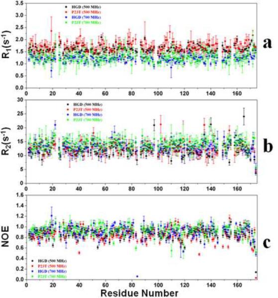
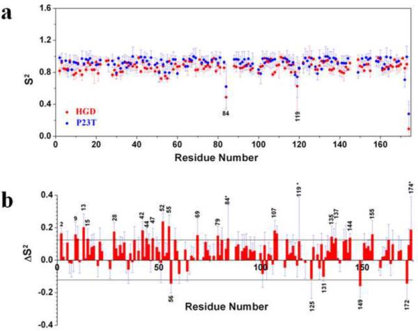
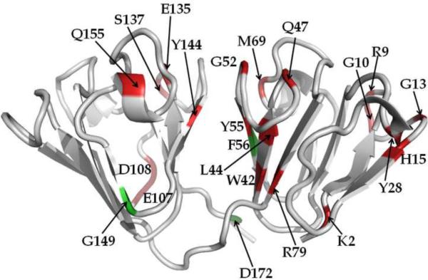
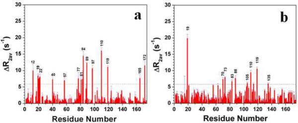
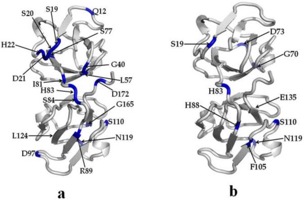
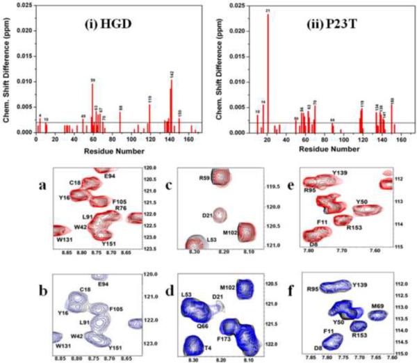
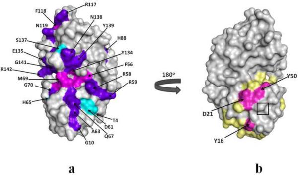
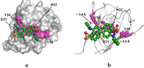
A number of point mutations in γD-crystallin are associated with human cataract. The Pro23-to-Thr (P23T) mutation is perhaps the most common, is geographically widespread, and presents itself in a variety of phenotypes. It is therefore important to understand the molecular basis of lens opacity due to this mutation. In our earlier studies, we noted that P23T shows retrograde and sharply lowered solubility, most likely due to the emergence of hydrophobic patches involved in protein aggregation. Binding of 4,4'-dianilino-1,1'-binaphthyl-5,5'-disulfonate (Bis-ANS) dye (a probe commonly used for detecting surface hydrophobicity) competed with aggregation, suggesting that the residues involved in Bis-ANS binding are also involved in protein aggregation. Here, using NMR spectroscopy in conjunction with Bis-ANS binding, we identify three residues (Y16, D21, and Y50) in P23T that are involved in binding the dye. Furthermore, using (15)N NMR relaxation experiments, we show that, in the mutant protein, backbone fluctuations are restricted to the picosecond-to-nanosecond and microsecond timescales relative to the wild type. Our present studies specify the residues involved in these two pivotal characteristics of the mutant protein, namely increased surface hydrophobicity and restricted mobility of the protein backbone, which can explain the nucleation and further propagation of protein aggregates. Thus, we have now identified the residues in the P23T mutant that give rise to novel hydrophobic surfaces, as well as those regions of the protein backbone where fluctuations in different timescales are restricted, providing a comprehensive understanding of how lens opacity could result from this mutation.


Figures

Fig. 1
Fig. 1
15N Relaxation parameters. (a) Backbone 15N longitudinal relaxation rates (R1) for HGD (black), P23T (red) at 500 MHz at pH 7.0, HGD (blue) and P23T (green) at 700 MHz at pH 7.0. (b) Backbone 15N transverse relaxation rates (R2) for HGD (black), P23T (red) at 500 MHz at pH 7.0, HGD (blue) and P23T (green) at 700 MHz at pH 7.0. (c) 1H-15N heteronuclear nOe values for HGD (black), P23T (red) at 500 MHz at pH 7.0, HGD (blue) and P23T (green) at 700 MHz at pH 7.0. Data were collected 25°C.
Fig. 2
Fig. 2
Model-free order parameter (S2) for P23T and HGD, and the difference in the order parameter between the two proteins (ΔS2). (a) S2 for HGD (red) and P23T (blue), as a function of residue number, at 25°C. (b) Per residue ΔS2 differences between P23T and HGD. ΔS2 are calculated as [data (mutant) - data (wild-type)]. Residues that show a ΔS2 value greater than the cutoff value of 0.125 were considered to be significant. Residues marked with an asterisk (*) are those for which the calculated errors are significantly higher than the average.
Fig. 3
Fig. 3
Differences in Model-free order parameters (ΔS2 from Fig. 2b) between HGD and P23T mapped on the crystal structure of HGD. Red represents residues for which backbone flexibility has been significantly reduced and green represents residues for which backbone flexibility has increased in P23T, relative to HGD.
Fig. 4
Fig. 4
CPMG data showing the residues which exhibit motion in the μs to ms time scale in HGD (a), and P23T (b). Data were collected at 700 MHz and at 25°C.
Fig. 5
Fig. 5
Cartoon representations of HGD and P23T highlighting the residues involved in slow dynamics (from Fig. 4). Residues showing slow motion (in μs to ms time scale) are marked with blue in HGD (a) and P23T (b), and are clearly reduced in P23T.
Fig. 6
Fig. 6
Upper panel: Amide chemical shift perturbations in the HSQC spectrum of HGD and P23T due to Bis-ANS binding. Shown here are average shifts in all the assigned residues in (1) HGD and (ii) P23T as a result of interaction with Bis-ANS. The weighted averages of the 1H and 15N chemical shifts perturbations are calculated as [(Δδ2NH + Δδ2N/25)/2]1/2. For the sake of clarity, all consecutive residues are unlabeled. Lower bottom panel: Overlay of HSQC spectra of free P23T (black contours) and Bis-ANS bound P23T (blue contours) showing interaction of (b) Y16, (d) D21 and (f) Y50 of P23T with Bis-ANS. The corresponding overlay of HSQC spectra of free HGD (lower top panel, black contours) and Bis-ANS bound HGD (red contours) are shown in (a), (c) and (e).
Fig. 7
Fig. 7
Molecular surface representation of HGD and P23T showing chemical shift perturbations as a result of interactions with Bis-ANS. (a) purple-blue indicates common residues showing interactions with Bis-ANS in both the proteins; cyan and magenta represent residues only in HGD and only in P23T, respectively, which interact with Bis-ANS. (b) A distinct surface patch (magenta) containing Y16, D21 and Y50 in P23T. The predicted hydrophobic patches (light yellow) are shown for comparison. The mutation site is shown in the boxed region in (b).
Fig. 8
Fig. 8
Bis-ANS docking on P23T. (a) Surface representation of P23T, showing Bis-ANS bound to the 4-residue (Y16, D21, H22 and Y50) hydrophobic patch. (b) Backbone cartoon representation of P23T-Bis-ANS complex describing the binding locus. Y16, D21, H22 and Y50 are shown in magenta.

Similar articles

Cited by

References

    1. Zetterstrom C, Lundvall A, Kugelberg M. Cataracts in children. J Cataract Refract Surg. 2005;31:824–40. - PubMed
    1. Hammond CJ, Snieder H, Spector TD, Gilbert CE. Genetic and environmental factors in age-related nuclear cataracts in monozygotic and dizygotic twins. N Engl J Med. 2000;342:1786–90. - PubMed
    1. Pande A, Pande J, Asherie N, Lomakin A, Ogun O, King JA, Lubsen NH, Walton D, Benedek GB. Molecular basis of a progressive juvenile-onset hereditary cataract. Proc Natl Acad Sci U S A. 2000;97:1993–8. - PMC - PubMed
    1. Pande A, Pande J, Asherie N, Lomakin A, Ogun O, King J, Benedek GB. Crystal cataracts: human genetic cataract caused by protein crystallization. Proc Natl Acad Sci U S A. 2001;98:6116–20. - PMC - PubMed
    1. Pande A, Annunziata O, Asherie N, Ogun O, Benedek GB, Pande J. Decrease in protein solubility and cataract formation caused by the Pro23 to Thr mutation in human gamma D-crystallin. Biochemistry. 2005;44:2491–500. - PubMed

Publication types

MeSH terms