. 2011 Aug 10;19(8):1127-37.
doi: 10.1016/j.str.2011.06.003.

Dynamics of the phosphoinositide 3-kinase p110δ interaction with p85α and membranes reveals aspects of regulation distinct from p110α

Affiliations

Dynamics of the phosphoinositide 3-kinase p110δ interaction with p85α and membranes reveals aspects of regulation distinct from p110α

John E Burke et al. Structure. .

Abstract

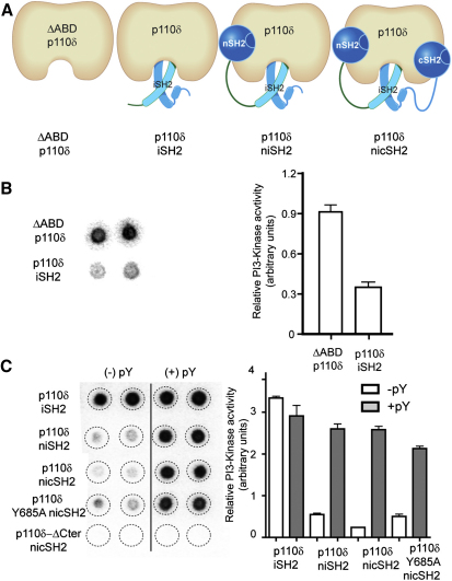
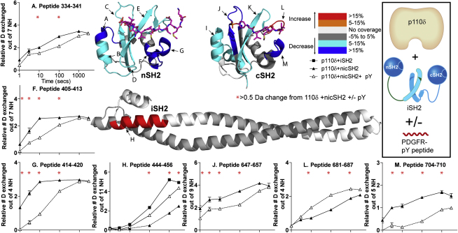
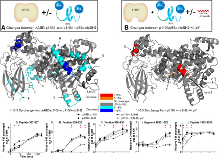
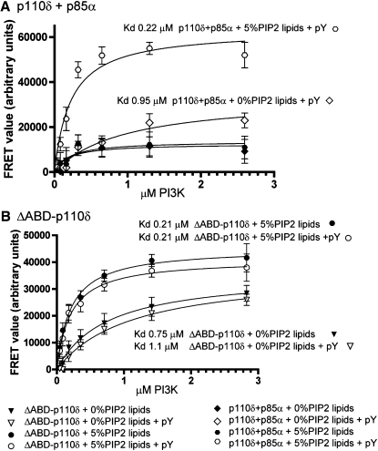
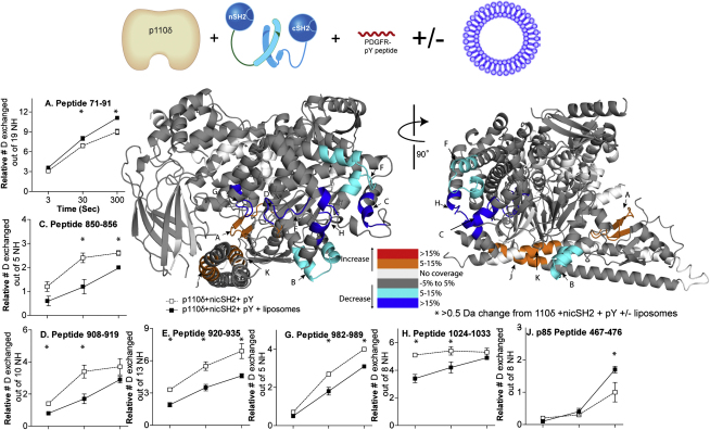
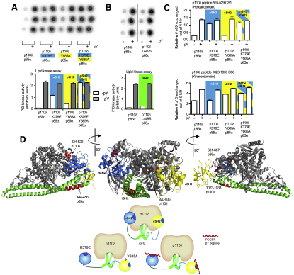
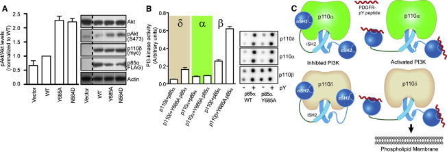
Phosphoinositide 3-kinase δ is upregulated in lymphocytic leukemias. Because the p85-regulatory subunit binds to any class IA subunit, it was assumed there is a single universal p85-mediated regulatory mechanism; however, we find isozyme-specific inhibition by p85α. Using deuterium exchange mass spectrometry (DXMS), we mapped regulatory interactions of p110δ with p85α. Both nSH2 and cSH2 domains of p85α contribute to full inhibition of p110δ, the nSH2 by contacting the helical domain and the cSH2 via the C terminus of p110δ. The cSH2 inhibits p110β and p110δ, but not p110α, implying that p110α is uniquely poised for oncogenic mutations. Binding RTK phosphopeptides disengages the SH2 domains, resulting in exposure of the catalytic subunit. We find that phosphopeptides greatly increase the affinity of the heterodimer for PIP2-containing membranes measured by FRET. DXMS identified regions decreasing exposure at membranes and also regions gaining exposure, indicating loosening of interactions within the heterodimer at membranes.


Figures

Figure 1
Figure 1
Kinase Activity of p110δ in the Presence of p85α Constructs and PDGFR pY (A) p110δ/p85α constructs tested for lipid kinase activity. (B) Kinase activity of ΔABD-p110δ compared with p110δ /iSH2 p85α. Assays measured 32P-PIP3 production in the presence of 0.5 nM enzyme, 100 μM ATP, and 5% PIP2 lipid vesicles at a concentration of 1 mg/ml. The left panel illustrates the autoradiogram of the filter (duplicate spots) and the right shows the quantitation of the spots. Kinase assays were performed in duplicate and repeated twice. The error bars show the standard deviation (SD). (C) In vitro kinase assay results for various p110δ and p85α constructs are shown. Assays measured 32P-PIP3 production in the presence of 5 nM enzyme, 100 μM ATP, and 5% PIP2 vesicles at a concentration of 1 mg/ml, +/− 10 μM PDGFR pY. Kinase assays were performed in duplicate and repeated twice.
Figure 2
Figure 2
Changes in Deuteration Levels of the p85α nicSH2 Construct (Bound to p110δ) in the Presence of 40 μM PDGFR pY Peptides spanning p85α (A–M) that showed >0.5 Da changes in deuteration level in the presence and absence of PDGFR pY for the p110δ+nicSH2 complex are mapped onto the structures (2VIY for the iSH2, 2IUI for the nSH2, and 1H9O for the cSH2). The percent change mapped on the structure according to the legend is the highest deuterium exchange difference change seen at any time point in the analysis. The structures of PDGFR pY bound to the nSH2 and cSH2 are colored purple. Only peptides that showed >10% change for more than two time points are graphed and shown below the structures. Experiments were performed in duplicate, and graphs are shown ± SD. All other peptides with changes >0.5 Da are shown in Figure S3. The graphs are labeled () for any time point with a >0.5 Da change for the p110δ+nicSH2 complex +/− PDGFR pY (see also Figure S3).
Figure 3
Figure 3
Changes in Deuteration Levels of p110δ Catalytic Subunit in the Presence of Both p85α and PDGFR pY (A) Peptides spanning p110δ (labeled A–I) that showed >0.5 Da changes in deuteration level in the presence and absence of the p85α nicSH2 are mapped onto the ΔABD-p110δ structure (2WXH) according to the legend. Peptides that showed >10% change for more than two time points are graphed and shown below the figure. Experiments were performed in duplicate, and graphs are shown ± SD. All other peptides with changes >0.5 Da are shown in Figure S4. (B) Peptides spanning the p110δ catalytic subunit in the p110δ+nicSH2 complex that showed >0.5 Da changes in deuteration level in the presence and absence of 40 μM PDGFR pY are mapped onto the structure. The percent change mapped on the structure according to the legend is the highest deuterium exchange difference change seen at any time point in the analysis. The area from 1020–1022 is named a segment to denote that this data was generated by subtraction of the deuterium level of peptide 1001–1019 from peptide 1001–1022. The graphs are labeled () for any time points with a >0.5 Da change between the ΔABD-p110δ and p110δ+nicSH2 constructs and () for any time point with a >0.5 Da change for the p110δ+nicSH2 +/− PDGFR pY (see also Figure S4).
Figure 4
Figure 4
Protein-Lipid FRET Measured from Intrinsic Tryptophanes to the DANSYL Probe of the DANSYL-PS-Containing Liposomes of the Free Catalytic Subunit and Full Length p110δ/p85α Complex in the Presence and Absence of PDGFR pY (A) Lipid binding of the p110δ/p85α complex with 0% and 5% PIP2 lipid vesicles in the presence and absence of PDGFR pY. (B) Lipid binding of the ΔABD-p110δ construct with 0% and 5% PIP2 lipid vesicles in the presence and absence of PDGFR pY. Experiments were repeated in triplicate and graphs are shown ± SD.
Figure 5
Figure 5
Changes in Deuteration Levels of p110δ and p85α in the Presence of 5% PIP2 Vesicles at 1 mg/ml A model for the iSH2 domain of p85α and ABD domain of the p110δ was generated by combining the ΔABD-p110δ structure (2WXH) with the structure of p110α in complex with niSH2 (3HIZ) (Mandelker et al., 2009). The C-terminal helix of the kinase domain that is disordered in p110δ is modeled (region H) from the structure of p110γ (1E7U) (Walker et al., 1999). Peptides spanning p110δ and p85α (labeled A–K) that showed >0.5 Da changes in deuteration level in the presence of vesicles are mapped onto the model. The percent change mapped on the structure according to the legend is the highest deuterium exchange difference change seen at any time point in the analysis. Peptides that showed a >10% change at any time point are graphed and shown below the figure. Experiments were performed in duplicate, and graphs are shown ± SD. All other peptides with changes >0.5 Da are shown in Figure S5. The graphs are labeled () for any time points with a >0.5 Da change between p110δ+nicSH2 + pY in the presence of lipids (see also Figure S5).
Figure 6
Figure 6
Effect of the p85α K379E and Y685A Mutations on Lipid Kinase Activity and Deuterium Exchange in Vitro with a Model of p110δ/p85α Regulatory Interaction (A) PI3K activity of p110δ, with full length p85α constructs containing nSH2 (K379E) and cSH2 (Y685A) mutations in the presence and absence of PDGFR pY. Assays measured 32P-PIP3 production in the presence of 10 nM enzyme, 100 μM ATP and 5% PIP2 lipid vesicles at a concentration of 1 mg/ml. PDGFR pY was 10 μM. (B) PI3K activity of p110δ, with full-length p85α containing the cancer-linked L449S iSH2 mutation in the presence and absence of PDGFR pY. All lipid kinase activity assays were performed in triplicate and graphs are shown ± SD. (C) The deuteration level at 1000 s of on-exchange for a helical domain peptide (524–529), and at 3 s of on exchange for a C-terminal peptide (1023–1033) was plotted for eight conditions as indicated on the legend. Experiments were performed in duplicate, and graphs are shown ± SD. (D) A structural model for the interaction of p110δ with the nSH2, iSH2, and cSH2 domains of p85α was generated using the crystal structure of the free p110δ catalytic subunit (2WXH), with the nSH2 from the p110α/p85α structure (3HHM), the iSH2 (2VIY), and the cSH2 from the recent p110β/p85β structure (2Y3A). Regions with changes on phosphopeptide binding are colored in red and labeled on the model (see also Figure S6).
Figure 7
Figure 7
Effect of the p85α Y685A Mutation on Lipid Kinase Activity In Vitro and PI3K Signaling in Cell Culture with a Model of p110δ/p85α Regulatory Interaction (A) Y685A mutation in the cSH2 of p85α (Y685A) increases Akt phosphorylation (Ser473) in HEK cells overexpressing p110δ+p85α heterodimers. Bar graphs show mean ± SEM (n = 3) of phosphorylated Akt (pAkt) to Akt ratios normalized to wild-type p110δ+p85α (WT). (B) PI3K activity of p110δ, p110α, and p110β in the presence of full length p85α (WT and Y685A) in the presence and absence of PDGFR pY. Assays measured 32P-PIP3 production in the presence of 10 nM enzyme, 100 μM ATP and 5% PIP2 lipid vesicles at a concentration of 1 mg/ml. PDGFR pY was 10 μM. PI3K assays were performed in duplicate and repeated three times. Shown is a representative experiment with graphs shown ± SD. (C) A model for the regulation of p110δ and p110α basal activity by the nicSH2 domains of p85α and activation by PDGFR pY. The presence of the nSH2 and cSH2 domains prevents lipid binding, and binding of PDGFR pY exposes lipid interacting regions and increases membrane affinity.

References

    1. Berndt A., Miller S., Williams O., Le D.D., Houseman B.T., Pacold J.I., Gorrec F., Hon W.C., Liu Y., Rommel C. The p110 delta structure: mechanisms for selectivity and potency of new PI(3)K inhibitors. Nat. Chem. Biol. 2010;6:117–124. - PMC - PubMed
    1. Burke J.E., Hsu Y.H., Deems R.A., Li S., Woods V.L., Jr., Dennis E.A. A phospholipid substrate molecule residing in the membrane surface mediates opening of the lid region in group IVA cytosolic phospholipase A2. J. Biol. Chem. 2008;283:31227–31236. - PMC - PubMed
    1. Burke J.E., Karbarz M.J., Deems R.A., Li S., Woods V.L., Jr., Dennis E.A. Interaction of group IA phospholipase A2 with metal ions and phospholipid vesicles probed with deuterium exchange mass spectrometry. Biochemistry. 2008;47:6451–6459. - PMC - PubMed
    1. Cantley L.C. The phosphoinositide 3-kinase pathway. Science. 2002;296:1655–1657. - PubMed
    1. Carson J.D., Van Aller G., Lehr R., Sinnamon R.H., Kirkpatrick R.B., Auger K.R., Dhanak D., Copeland R.A., Gontarek R.R., Tummino P.J., Luo L. Effects of oncogenic p110alpha subunit mutations on the lipid kinase activity of phosphoinositide 3-kinase. Biochem. J. 2008;409:519–524. - PubMed

Publication types

MeSH terms

Substances