

Image similarity and tissue overlaps as surrogates for image registration accuracy: widely used but unreliable
- PMID: 21827972
- PMCID: PMC3274625
- DOI: 10.1109/TMI.2011.2163944
Image similarity and tissue overlaps as surrogates for image registration accuracy: widely used but unreliable
Abstract
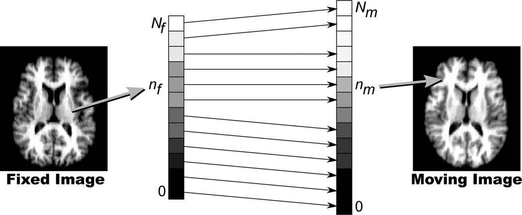
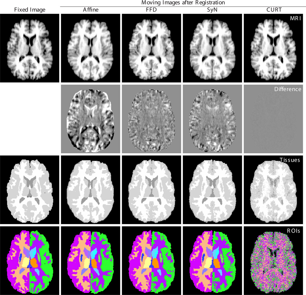
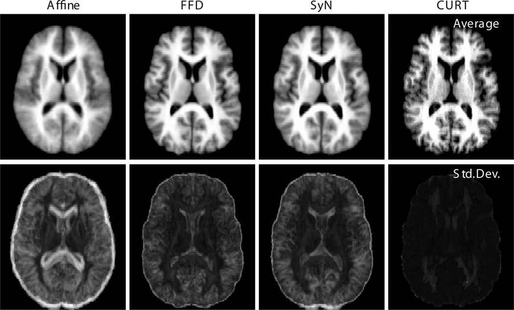
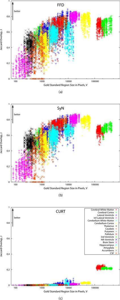
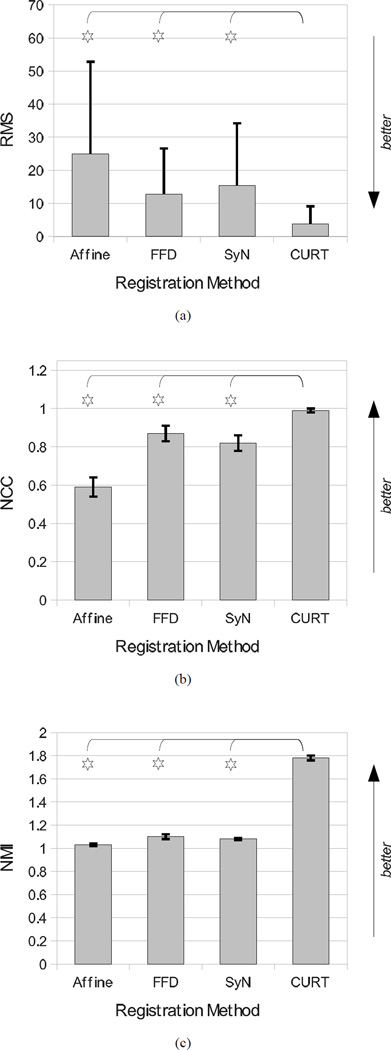
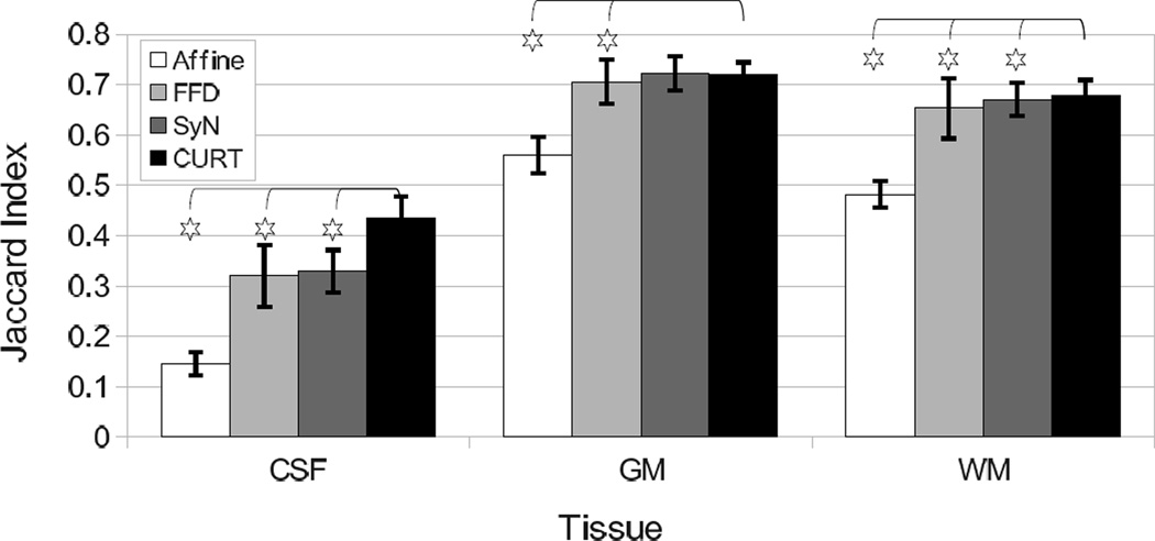
The accuracy of nonrigid image registrations is commonly approximated using surrogate measures such as tissue label overlap scores, image similarity, image difference, or transformation inverse consistency error. This paper provides experimental evidence that these measures, even when used in combination, cannot distinguish accurate from inaccurate registrations. To this end, we introduce a "registration" algorithm that generates highly inaccurate image transformations, yet performs extremely well in terms of the surrogate measures. Of the tested criteria, only overlap scores of localized anatomical regions reliably distinguish reasonable from inaccurate registrations, whereas image similarity and tissue overlap do not. We conclude that tissue overlap and image similarity, whether used alone or together, do not provide valid evidence for accurate registrations and should thus not be reported or accepted as such.
Figures
References
-
- West JB, Fitzpatrick JM, Wang MY, Dawant BM, Maurer CR, Jr, Kessler RM, Maciunas RJ, Barillot C, Lemoine D, Collignon A, Maes F, Suetens P, Vandermeulen D, van den Elsen PA, Napel S, Sumanaweera TS, Harkness B, Hemler PF, Hill DLG, Hawkes DJ, Studholme C, Maintz JBA, Viergever MA, Malandain G, Pennec X, Noz ME, Maguire GQ, Jr, Pollack M, Pelizzari CA, Robb RA, Hanson D, Woods RP. Comparison and evaluation of retrospective intermodality brain image registration techniques. J. Comput. Assist. Tomogr. 1997;vol. 21(no. 4):554–566. - PubMed
-
- Murphy K, van Ginneken B, Klein S, Staring M, de Hoop B, Viergever M, Pluim J. Semi-automatic construction of reference standards for evaluation of image registration. Med. Image. Anal. 2011 Feb.vol. 15(no. 1):71–84. - PubMed
-
- Fitzpatrick JM, West JB, Maurer CR., Jr Predicting error in rigid-body, point-based registration. IEEE Trans. Med. Imag. 1998 Oct.vol. 17(no. 5):694–702. - PubMed
-
- Song JH, Christensen GE, Hawley JA, Wei Y, Kuhl JG. Evaluating image registration using NIREP. In: Fischer B, Dawant BM, Lorenz C, editors. Biomedical; Image Registration — 4th International Workshop, WBIR 2010; July 11–13, 2010; Lübeck, Germany. Berlin/Heidelberg: Springer-Verlag; 2010. pp. 140–150. Proceedings, ser. LNCS.
-
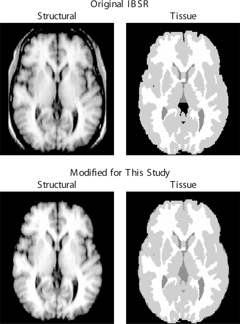
- Internet Brain Segmentation Repository (IBSR) [Online]. Available: http://www.cma.mgh.harvard.edu/ibsr/
Publication types
MeSH terms
Substances
Grants and funding
LinkOut - more resources
Full Text Sources
Other Literature Sources
Medical
