. 2011 Sep 30;286(39):34184-98.
doi: 10.1074/jbc.M111.247783. Epub 2011 Aug 2.

Gelsolin and non-muscle myosin IIA interact to mediate calcium-regulated collagen phagocytosis

Affiliations

Gelsolin and non-muscle myosin IIA interact to mediate calcium-regulated collagen phagocytosis

Pamma D Arora et al. J Biol Chem. .

Abstract

The formation of adhesion complexes is the rate-limiting step for collagen phagocytosis by fibroblasts, but the role of Ca(2+) and the potential interactions of actin-binding proteins in regulating collagen phagocytosis are not well defined. We found that the binding of collagen beads to fibroblasts was temporally and spatially associated with actin assembly at nascent phagosomes, which was absent in gelsolin null cells. Analysis of tryptic digests isolated from gelsolin immunoprecipitates indicated that non-muscle (NM) myosin IIA may bind to gelsolin. Immunostaining and immunoprecipitation showed that gelsolin and NM myosin IIA associated at collagen adhesion sites. Gelsolin and NM myosin IIA were both required for collagen binding and internalization. Collagen binding to cells initiated a prolonged increase of [Ca(2+)](i), which was absent in cells null for gelsolin or NM myosin IIA. Collagen bead-induced increases of [Ca(2+)](i) were associated with phosphorylation of the myosin light chain, which was dependent on gelsolin. NM myosin IIA filament assembly, which was dependent on myosin light chain phosphorylation and increased [Ca(2+)](i), also required gelsolin. Ionomycin-induced increases of [Ca(2+)](i) overcame the block of myosin filament assembly in gelsolin null cells. We conclude that gelsolin and NM myosin IIA interact at collagen adhesion sites to enable NM myosin IIA filament assembly and localized, Ca(2+)-dependent remodeling of actin at the nascent phagosome and that these steps are required for collagen phagocytosis.


Figures
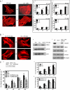
FIGURE 1.
FIGURE 1.
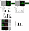
A, a, gelsolin WT and gelsolin null cells with and without treatment with JA (500 nm) and stained with rhodamine-phalloidin. b, gelsolin WT and null cells were treated with JA and plated on collagen-coated beads. Data are means ± S.E. of bound collagen beads per cell (n = 100 cells/group). *, difference of p < 0.05. c, similarly, the % bead internalization was not different in the presence or absence of JA in gelsolin null cells and gelsolin WT cells. B, a, phalloidin-stained cells showed no difference in actin filament distribution in untreated or JA-treated cells. b, total cell lysates from NMIIB null, NMIIA null, and WT ES cells were immunoblotted for the indicated proteins. c, similarly, cell lysates from gelsolin WT, gelsolin null cells, and gelsolin null cells transfected with gelsolin cDNA show levels of indicated proteins. C, a, fibroblasts from gelsolin null mice transfected with NMIIA siRNA show >70% reduction of NMIIA protein levels compared with cells treated with non-targeted siRNA. b, the numbers of collagen beads binding per cell were significantly (p < 0.05) lower in gelsolin null cells and in gelsolin null cells transfected with siRNA NMIIA. c, in fibroblasts from WT mice treated with NMIIA or non-targeted siRNA as control, internalized beads were discriminated by quenching the fluorescence of extracellular bound beads with trypan blue. Fluorescent and quenched beads were counted in 60 cells/sample at each time point, and NMIIA knockdown cells showed reduced internalization for all time points (p < 0.02). d, collagen bead internalization assays on gelsolin WT and null cells.
FIGURE 2.
FIGURE 2.
A, recruitment of F-actin (GFP-Lifeact) to bead binding sites imaged by TIRF microscopy in living gelsolin WT (a) and gelsolin null cells (b). Insets show fluorescence at the collagen-coated bead sites in gelsolin WT and null cells. DIC, differential interference contrast. B, a, WT and gelsolin null cells were co-transfected with RFP-Lifeact and GFP-NMIIA. Images were taken on live cells over 60 min. Histograms show mean fluorescence ± S.E. from regions of interest (3-μm diameter circles) around individual bead sites obtained from the images of 30 different cells at each time point. b, line graph shows the mean ± S.E. of the ratios of RFP-Lifeact to GFP-NMIIA fluorescence from around beads in 30 different cells for each time point. C, representative images of NMIIA WT, NMIIA B null, and NMIIA null cells showing the incorporation of rhodamine-labeled actin monomers in permeabilized cells to estimate free barbed ends. Cells were stained with Alexa 488 phalloidin to show F-actin. Histogram shows means ± S.E. of rhodamine fluorescence from 50 cells for each condition.
FIGURE 3.
FIGURE 3.
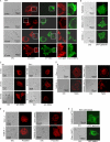
A, representative images showing distribution of GFP-NMIIA and endogenous gelsolin in WT cells. Insets show the targeting of gelsolin and NMIIA proteins to the developing phagosomes. B, NMIIB null, NMIIA WT, or non-muscle myosin IIA-deficient ES cells showing localization of GFP-gelsolin to collagen-coated beads after 20 min of incubation. A total of 20 cells were examined. These experiments were repeated three times. C, collagen-coated beads incubated with gelsolin WT and gelsolin null cells show enhanced localization of endogenous NMIIA to beads over time. D, a and b, gelsolin WT and gelsolin null cells and NMIIA WT ES cells and NMIIA null ES cells show an equivalent accumulation of talin and paxillin at collagen beads by immunostaining for talin (a) and paxillin (b). c, BSA-coated beads incubated with WT NMIIA and WT gelsolin cells transfected with GFP gelsolin and GFP NMIIA, respectively. A total of 25 cells were examined. DIC, differential interference contrast.
FIGURE 4.
FIGURE 4.
A, a and b, NMIIA or gelsolin immunoprecipitates of bead-associated proteins were immunoblotted for gelsolin and NMIIA, respectively. In separate experiments an irrelevant antibody was used for immunoprecipitation, and immunoprecipitates were immunoblotted with NMIIA and gelsolin. c, gelsolin immunoprecipitates of bead-associated proteins were immunoblotted for NMIIA in response to BSA-coated beads in the presence or absence of Ca2+. B, gelsolin null cells transfected with FLAG-tagged G1–G6, G1–G3, and G4–G6 constructs were incubated with collagen beads for 30 min. Bead-associated proteins immunoprecipitated (IP) with FLAG antibody were immunoblotted (WB, Western blot) for NMIIA (a). TCL, total cell lysates (b). These observations were made in four different experiments. C, gelsolin null cells transfected with FLAG-tagged G1–G6 and FLAG-tagged empty construct. Cells were incubated with collagen beads for 30 min, and bead-associated proteins were immunoprecipitated with FLAG antibody and then immunoblotted for NMIIB.
FIGURE 5.
FIGURE 5.
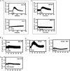
A, fura2/AM-loaded gelsolin (Gsn) WT and gelsolin null cells (a) and NMIIA WT and NMIIA null cells (b) showing Ca2+ fluxes in response to collagen-coated bead (CCB) binding measured by ratio fluorimetry. B, a and b, Ca2+ fluxes in response to collagen bead binding in gelsolin WT and null cells treated with JA (500 nm) and latrunculin B (LatB, 1 μm), respectively. c, intracellular calcium ion responses to collagen-coated beads in latrunculin B (1 μm)-treated NMIIA−/− cells. d, calcium responses were also measured in gelsolin WT cells after incubation with BSA-coated beads as controls. Data are means ± S.E. of bound collagen beads per cell (n = 100 cells/group).
FIGURE 6.
FIGURE 6.
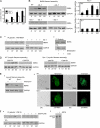
A, cells treated with ionomycin exhibit myosin light chain phosphorylation in WT and gelsolin null cells. B, a, bead-associated proteins collected from cells treated with collagen-coated beads show maximal phosphorylation of MLC at 30 min in WT cells but not in gelsolin-deficient cells. The histogram below shows ratios of quantification of blot densities as indicated. Data are mean ± S.E. of ratios. b, immunostaining shows localization of phospho-MLC (pMLC) at the bead sites after 10 and 30 min incubation with collagen-coated beads. C, in the presence of the MLC kinase inhibitor ML-7, gelsolin WT or null cells incubated with collagen beads show a complete block of MLC phosphorylation. The experiment was repeated three times. DIC, differential interference contrast; CCB, collagen-coated beads; tMLC, total myosin light chain.
FIGURE 7.
FIGURE 7.
A, gelsolin WT cells treated with ML-7 (25 μm) show reduced collagen binding (*, p < 0. 01 after 10 min). Data are mean ± S.E. numbers of collagen beads bound to cells (n = 75 cells/group). B, ML-7 treated or untreated gelsolin WT and null cells were incubated with collagen beads and probed with NMIIA antibody. Histograms are the ratios of quantification of blot densities as indicated. Data are mean ± S.E. of ratios. C, gelsolin WT cells treated with or without ML-7 (a) and with or without blebbistatin (b). Gelsolin immunoprecipitates (IP) from collagen-treated samples were probed with NMIIA antibody. CCB, collagen-coated beads; TCL, total cell lysates; WB, Western blot. D, cells loaded with the Ca2+ chelator BAPTA/AM and incubated with collagen beads inhibit myosin filament assembly in Triton-insoluble fractionation assay. E, a, ionomycin treatment of gelsolin null cells overcomes the block of myosin filament assembly in Triton-insoluble fractionation assay. b, ionomycin-treated gelsolin null cells show targeting of NMIIA to bead sites in collagen binding assays. DIC, differential interference contrast. F, a, in the presence or absence of BAPTA/AM, gelsolin immunoprecipitates after incubation with beads collagen were probed with NMIIA antibody. b, GST-Sepharose beads with bound gelsolin and NMIIA (residues 1338–1960) at a 2:1 molar ratio were incubated for 1 h in Ca2+- or EGTA-containing buffer followed by extensive washing. Bead-associated proteins were eluted and separated by SDS-PAGE. Gelsolin associated with NMIIA in the presence of Ca2+ but not in the presence of EGTA.
FIGURE 8.
FIGURE 8.
Schematic diagram indicating the functional dependence of collagen phagocytosis on NMIIA interactions with gelsolin. 1, initial binding of collagen to β1 integrin at collagen bead adhesion sites. 2a, localized Ca2+ flux enhances phosphorylation of myosin light chain at serine 19 and increases NMIIA filament assembly. 2b, Ca2+ flux induces activation of gelsolin and MLCK, myosin light chain kinase. 3, gelsolin is required for MLC phosphorylation because the absence of [Ca2+]i in gelsolin null cells prevents the phosphorylation of NMIIA MLC required for myosin IIA filament assembly. 4, NMIIA filaments interact with gelsolin to provide anchorage for gelsolin at collagen bead binding sites. 5, NMIIA filaments provide Rap1 localization at bead sites and enable β1 integrin activation (12). 6, spatial localization of gelsolin by NMIIA filaments provides remodeling of actin filaments around bead binding sites, which is required for phagosome formation.

Similar articles

Cited by

References

    1. Pérez-Tamayo R. (1978) Am. J. Pathol. 92, 508–566 - PMC - PubMed
    1. Murphy G., Nagase H. (2008) Mol. Aspects Med. 29, 290–308 - PMC - PubMed
    1. Caron E., Hall A. (1998) Science 282, 1717–1721 - PubMed
    1. Krause K. H. (2000) Schweiz. Med. Wochenschr. 130, 97–100 - PubMed
    1. Everts V., van der Zee E., Creemers L., Beertsen W. (1996) Histochem J. 28, 229–245 - PubMed

Publication types

MeSH terms